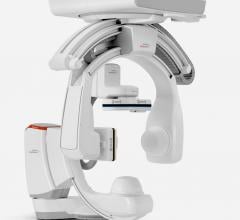
Over the last 10 years, hospitals have phased out analog image intensifiers for flat-panel, digital detector angiography systems in their cath labs. As hospitals look to purchase current-generation digital angiography systems, buyers should be aware of several new technologies.
Better Navigation Aids A key feature of newer digital angiography systems is the use of rotational angiography, in which the C-arm makes a quick rotation around the patient to create a computed tomography (CT)-like 3-D image. A CT image has a resolution of about 1 Hounsfield unit (HU), while the resolution of a CT-like angiography image is about 10 HU.(1) These dataset images can be used to segment out vessels or anatomy of interest as a navigation aid. Most systems also allow overlays on these CT-like images on the fluoroscopic view to help better define structures and vessels to aid navigation and reduce the amount of contrast. These roadmap images can be fused with the orientation of the C-arm for better catheter navigation. Today’s angiography systems offer software to take CT or magnetic resonance imaging (MRI) datasets that can be registered with the fluoro view. These hybrid images can then be rotated with the orientation of the C-arm to help with navigation. The opacity of the overlay image can be adjusted for optimal viewing of the live angio. Some systems also offer software to enhance percutaneous devices. These features help identify if stents are fully apposed to the vessel wall, or to better visualize the results of bifurcation stenting. An example of this is GE Healthcare’s StentVis, which uses elastic registration that takes radiopaque markers and guide wires into account to help create more contrast to better visualize stents.
Dose-Lowering Technologies Manufacturers developed a variety of new features to help lower and monitor ionizing X-ray radiation dose. Features include image enhancement software to enable the use of lower-dose imaging and easy table-side frame rate adjustment. An example of this is Toshiba’s Infinix-i, where tableside controls can adjust the pulse fluoro frame rate settings. The system also offers virtual collimation, where an electronic outline guides collimator blade placement, avoiding the use of fluoroscopy for positioning. GE’s Dynamic Range Management (DRM) software enhances structures of interest on hard-to-read angiographic images to transform background thickness, vessel contrast, background contrast, background brightness and reduce problems with image blackout and burnout. Siemens Healthcare’s Artis zee system offers several Combined Applications to Reduce Exposure (CARE) modules to assist with dose reduction without compromising image quality. CARE Vison uses low pulsing frequencies to meet individual dose-saving targets below 10 p/s, down to 0.5 p/s. CARE Filter uses additional Cu-filters to reduce skin dose by more than 70 percent in some patients based on patient weight. CARE Profile adjusts the collimators and wedge filters without using fluoroscopy. Some vendors offer dose monitoring to warn clinicians when they enter the radiation field (Philips’ DoseAware) or track radiation usage (GE’s Innova Dose Reports).
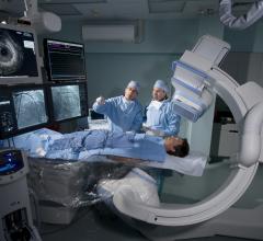
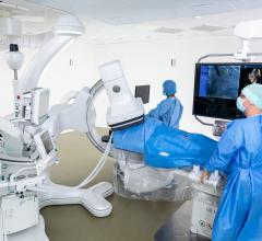
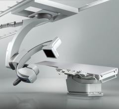
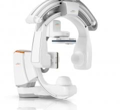
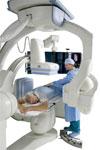
Hybrid OR Needs Minimally invasive and transcatheter procedures continue to reduce the need for open surgery, which has placed greater demands for angiography systems in the surgical suite. Also, many hospitals are developing hybrid operating rooms (ORs) so both surgical and interventional procedures can be performed in one location. If considering a hybrid OR, key angiography system considerations include installing displays that can easily be sterilized, the ability of the C-arm to be moved out of the way for better patient access and the movement of the patient table. Surgical tables need to be raised and lowered for better patient accessibility. Some tables also can tilt to raise different parts of the body to help with surgical access. Angiography systems come in both floor- and ceiling-mounted configurations, which may be a deciding factor in some installations. ORs require much larger square footage than cath labs to accommodate additional staff, anesthesiology, surgical equipment and space to transfer patients. Some hospitals have found ceiling-mounted systems are useful to swing the C-arm completely out of the way and to eliminate obstacles on the floor, especially in smaller hybrid suites.
Interface Considerations Angiography systems are no longer used as a sole imaging or diagnostic modality in the cath lab, so interfaces with other systems help improve workflow. Ultrasound is playing an increasing role, with intravascular ultrasound (IVUS) to guide vascular interventions and intracardiac echo (ICE) and transesophageal echo (TEE) to help guide structural heart procedures. Conventional ultrasound systems are also seeing increased use to reduce X-ray radiation and guide procedures, especially in peripheral vascular interventions. Optical coherence tomography (OCT) and fractional flow reserve (FFR) also are becoming key tools in guiding vascular interventions. Electrophysiology (EP) frequently shares the cath lab, so integration with EP mapping and navigation systems also might be an important consideration. References: 1. Jacques Kpodonu. “Manual of Thoracic Endoarortic Surgery.” Springer, 2010.
 January 31, 2024