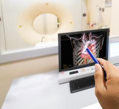
This is a 3-D view of blood flow through the chambers of a fetal heart using the GE Healthcare fetalHQ analysis software.
Many of the latest advances in cardiovascular imaging technologies are unveiled each year at the Radiological Society of North America (RSNA) meeting. This is the world's largest radiology conference, held each year in Chicago the week after Thanksgiving in November. At RSNA 2018, there were several new innovations in cardiac imaging released.
Cardiac Ultrasound Software Streamlines Fetal Heart Exams
Detailed fetal cardiac assessments are difficult to perform because of the small size and extremely fast heart rates. At 18 weeks, the fetal heart is the size of an olive and beating about 150 times per minute. Additionally, the structure itself is extremely complex and with the baby in constant motion, it is always a moving target. Imaging is important, because congenital heart defects affect one out of every 110 babies born globally.
Offering a potential solution to aid fetal evaluations, at RSNA, GE Healthcare unveiled its fetalHQ heart and vascular software for fetal ultrasound. The software for the Voluson E10 helps evaluate the fetal heart shape, size and contractibility in less than 3 minutes. A feature called Radiant Flow shows the blood flow in a 3-D view. It can also help show slow-flow blood, such as neurovascular circulation.
Watch a VIDEO example of this technology showing blood flow and Radiant Flow.
Watch a VIDEO showing this technology tracking fetal heart contractibility.
This technology was developed after Greggory DeVore, M.D., a specialist in maternal fetal medicine at Huntington Hospital, Pasadena, Calif., modified adult echo speckle tracking analysis software to look at the fetal heart. DeVore reprogrammed the software to divide the fetal heart in 24 segments, measuring the heart in ways that had never been done before.
“This is an incredible step forward in examining the fetal heart,” DeVore explained. “Previously, we had various tools that looked at specific sites of the heart, but nothing that examined the entire chamber.” On top of this, the heart had to be in a specific position — at 12 or six o’clock on the screen to get the proper measurements. And this all required an advanced level of diagnostic skills to learn the techniques.
For example, coarctation of the aorta is a narrowing of the aorta, the large blood vessel that branches off the heart and delivers oxygen-rich blood to the body. When this occurs, the heart must pump harder to force blood through the narrowed part of the aorta. When examining the fetal heart’s anatomy, there are findings associated with coarctation, but are not always 100 percent specific for this condition. Because of this, the clinician often has to play a guessing game to decide whether coarctation is present or not. By looking at the function rather than the structure with fetalHQ, DeVore found he was able to separate these cases with almost 100 percent accuracy.
In addition, DeVore described cases where the fetus was not growing properly. Typically, this is defined as a weight less than the tenth percentile, and then divided into three groups. One of these groups entails fetuses that are between the third and tenth percentile with normal blood flow to the placenta and brain. While small, these pregnancies are typically deemed “normal” with no risk factors.
However, when DeVore examined 50 of these cases with fetalHQ, he discovered abnormal cardiac function. “This changes the whole paradigm as to how you interpret and manage these types of fetuses going forward,” DeVore said. “It allows us to ask, ‘How is the heart working?’ We’re now able to identify abnormal function, shape and size of the heart – things we couldn’t see previously.”
Cardiac Echo Exams Via an iPhone
Point-of-care ultrasound recently got a major technology boost with the introduction of the Butterfly IQ ultrasound transducer and app. The company displayed at RSNA for the first time in 2018, and showed attendees how they can convert their iPhones into a reasonably good quality diagnostic ultrasound system. Their U.S. Food and Drug Administration (FDA)-cleared technology is the first “ultrasound system on a chip” to be released commercially. It consists of a transducer that connects to an iPhone or iPad to record ultrasounds.
The system has 18 different applications for specialized images, including cardiac imaging, vascular, aorta, lung, abdominal and others. The apps allow for quantification and offer features usually found only on larger cart-based systems. The company said 90 percent of the work that can be done on a cart-based system can be performed with the Butterfly IQ. The apps allow basic measurements and annotations, and images can be transferred from the device into a cloud server for storage or transferring to a picture archiving and communication system (PACS) as a DICOM format. The images also can be saved as a PNG or videos in the MP4 format.
The ultrasound system on a chip technology allows a single transducer to be reconfigured with a simple setting to image as a curved, phased or linear probe, rather than needing to physically have and swap out various transducers. The system also allows for changes on the fly for beam sharpness, frequency and focal depth. The system also offers there scanning modes — M-mode, B-mode and color Doppler. The 2-D Butterfly transducer is composed of 9,000 elements and does not use traditional Piezo crystal technology.
Leveraging the fact that the platform operates on a smartphone, users can tag colleagues on to images they create to get quick consultations in or outside of the hospital. The tags create a de-identified image link so there is no violation of HIPAA privacy regulations.
This technology was picked as one of the big ultrasound advances in the VIDEO: 4 Recent Advances in Echocardiography Technology.
Introduction of a New Type of SPECT-CT Scanner
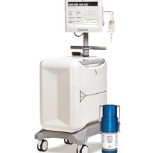
Spectrum Dynamics introduced a really interesting new single photon emission computed tomography CT (SPECT-CT) scanner at RSNA 2018. The FDA-cleared Veriton imaging system uses 12 SPECT detectors composed of 96 elements mounted on robotic arms. The arms automatically move toward the patient and use a sensor to stop a few millimeters from the skin to optimize photon counts, allow focusing on specific organs and to improve overall SPECT image quality. It also uses more sensitive CZT digital detectors, which allows either faster scan times, or use of only half the radiotracer dose of analog detector scans. The company said the images produced are twice the resolution of conventional SPECT cameras.
The CT system offers a large 80 cm bore and comes in 16-, 64- or 128-slice configurations. It is field upgradeable to higher slice CT.
Four systems are currently installed in France and the company was hoping to have its first U.S. install completed by the summer of 2019.
Watch the VIDEO: Walk Around of the Veriton SPECT-CT System.
Virtual Contrast Angiography Using AI
GE Healthcare showed its new Liver Assist Virtual Injection (VI) technology it developed for interventional radiology. It incorporates applied intelligence-enhanced software that identified the feeder blood vessels for liver tumors and identified the best locations to embolize the tumor. The AI also can enhance the blood to look like a contrast injection in the interventional lab to help map the vessels for procedural pre-planning prior to having the patient on the table. The software can show the direction of flow in each vessel segment and can overlay a vessel map onto 3-D rotational angiography images created in the cath lab to aid procedural navigation.
Watch the VIDEO: Editor's Choice of the Most Innovative New Imaging Technologies at RSNA 2018


 March 05, 2024