The Philips Azurion angiography system offers several imaging and software enhancements to aid guidance during complex interventional procedures.
Five new angiography systems were launched in 2017 that included improvements to speed workflow and improve both image quality and interventional procedural guidance.
Two of the biggest advances in angiography last year were the release of the new Siemens Artis Pheno and Philips Azurion systems, which both gained U.S. Food and Drug Administration (FDA) clearance in early 2017, said MD Buyline Cardiology Market Analyst Tom Watson. “The Philips Azurion is really focused on adding workflow features that help the physician without having to go to multiple views, imaging systems or screens. The system consolidates that data into one place. It enables physicians to be able to make decisions very quickly and on the fly.”
He said Siemens' new Pheno system was developed to address issues raised by end-users of the vendor’s Artis Zeego system. “The Pheno adds features and capabilities to aid the workflow,” Watson said. “They are adding a more robust table for larger patients.”
In addition to those two systems, Shimadzu and Canon Medical Systems (formerly Toshiba) launched sales of two new systems. Siemens also unveiled a combined computed tomography (CT)/magnetic resonance imaging (MRI) angiography system at the 2017 Radiological Society of North America (RSNA) annual meeting.
Shimadzu’s Trinias Unity
Shimadzu Medical Systems introduced its Trinias Unity interventional X-ray system at the 2017 Transcatheter Cardiovascular Therapeutics (TCT) meeting. It incorporates technologies to speed and automate workflow, and a new patient table with more positioning possibilities than previous systems. A new feature is a smart-touch display attached to the table-side controls. It allows users control for all the table-side acquisitions and to customize the configuration of the screens on the overhead monitor.
For cardiology, the Trinias comes in 8- and 12-inch detector sizes. A larger 12 x 16-inch detector was introduced at RSNA 2017, targeted for vascular and interventional radiology applications.
Two features operators may find useful are the Real-Time Smooth Mass (RSM) subtraction and Stentview. RSM cleans up the image and compensates for motion blur if the patient moves. Stentview offers real-time stent visualization, so the stent can be visualized while it is being expanded or post-inflation on live fluoro, rather than as a post-processed image.
Canon Infinix-i Sky +
At RSNA 2016, Canon Medical Systems introduced its newest angiography system, the Infinix-i Sky +. The company is promoting the system as enabling 3-D imaging anywhere in the anatomy with its unique sliding double C-arm design to deliver a large amount of flexibility. Its C-arm flip and 3-D imaging can image patients from head-to-end and left or right side. This flexibility is aimed primarily at advanced interventional radiology and oncology procedures. The system uses a ceiling-mounted gantry with more capabilities than previous Canon ceiling-mounted systems. This includes much faster rotational angiography 3-D image acquisitions. It also allows the system to be pulled out of the way and parked in a corner of the room for conversion to open surgical procedures or to aid patient transfers. It comes with a standard 12 x 16-inch detector.
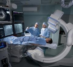
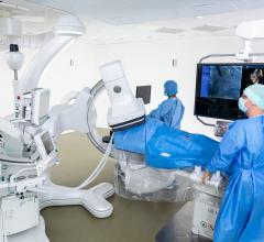
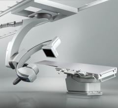
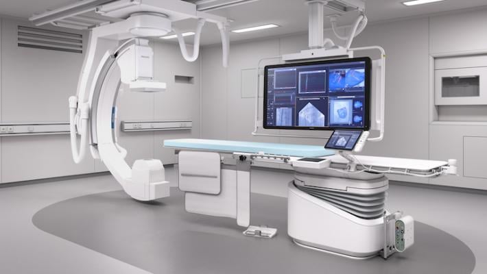
Philips’ Azurion
Philips began its global launch of the new Azurion system in early 2017. The vendor said it is its next-generation image-guided therapy platform.
The company said the platform features a state-of-the-art ergonomic design with an easy-to-use intuitive user interface, enabling clinicians to swiftly and confidently perform a wide range of routine and complex procedures in the interventional lab. With the rapid growth of image-guided therapy procedures, hospitals and health systems are increasingly faced with the need to control costs, while improving their standards of care. Philips said Azurion has been designed to address these challenges and is equipped with new workflow options and performance dashboards, as well as an upcoming range of productivity improvement services.
The Azurion platform features over 1,000 new components, including an enhanced flat-panel detector, and Philips’ newly developed ConnectOS operating system for the seamless integration of real-time information from all relevant technologies in the interventional lab. All of these components work together to deliver high image quality at ultra-low X-ray dose and real-time image processing on multiple work spots within the interventional lab. Parallel working enables the team of clinicians to complete different tasks simultaneously in the interventional lab, saving valuable time without compromising quality of care. Azurion features procedure cards that allow clinicians to pre-program routine tasks and user preferences, helping to minimize preparation errors and further reducing preparation and procedure time.
Advanced interventional tools integrated into the Azurion platform include VesselNavigator for vascular surgery; EchoNavigator for interventional cardiology; EmboGuide for inteventional oncology; and AneurysmFlow for interventional neuroradiology.
In July 2017, Philips gained FDA clearance for a couple additional navigation technologies for its Azurion system, including Dynamic Coronary Roadmap, which uses a contrast cine run to create a red vessel overlay on live fluoro to aid navigation. This can reduce the need for contrast because the vasculature can already be seen on the screen. The technology is also motion compensated to help stabilize the fused images to better visualize the catheters.
The second new technology is StentBoost Live, which freezes motion to get a better view of stents. It helps visualize small manipulations of the stent. The technology is an improved version of the previously available Stent Boost software.
At RSNA 2017, Philips released results for an independent, two-year study demonstrating the clinical workflow benefits of the Azurion. The study investigated nearly 800 patient procedures to evaluate the impact of Azurion at St. Antonius Hospital in Nieuwegein, the Netherlands. The data demonstrated a 17 percent reduction of the average interventional procedure time, a 12 percent reduction of in-lab patient preparation time and a 28 percent reduction of post-procedure lab time.
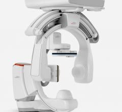
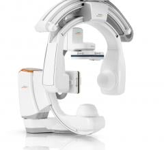
Siemens’ Artis pheno
The U.S. Food and Drug Administration (FDA) cleared the Siemens Healthineers Artis pheno robotic C-arm angiography system in March 2017. It reduces the amount of cabling often used in these systems, boasts an antimicrobial coating to help with infection control, and can fold up and park in a corner of the room for hybrid labs, or for increased patient access. The system has a CPR button that immediately drops the table to the floor and automatically swings the C-arm out of the way to better facilitate resuscitation efforts. The weight of the patient table was also increased to 617 pounds. Another design feature to accommodate obese patients is a larger space between the detector and the X-ray source to facilitate rotational angiography. Another unique feature is the system comes integrated with a Freestyle wireless ultrasound system. The ultrasound system is installed on the back of the main display screen and the wireless transducer can be used anywhere in the room. The system is particularly helpful for percutaneous vessel access, anesthesiology and needle biopsies.
The pheno also possesses a new 2k recording technology capable of delivering 2-D imaging resolution four times higher in all recording processes than its predecessor system, the Artis zeego.
The StructureScout exposure control feature can regulate acquisition parameters to automatically achieve optimal image contrast at ALARA (As Low As Reasonably Achievable) dose levels. And because the system also can scan up to 68 percent faster than its predecessor, its syngo DynaCT clinical software application can produce 3-D images that use less contrast media, increasing patient safety by decreasing the load on the kidneys.
Software applications to aid users in performing a variety of interventional procedures include CLEARstent Live for real-time verification of stent positioning during implantation; syngo CTO Guidance for better clinician planning by delivering automatic segmentation of coronary computed tomography angiograms (CCTAs) as well as providing procedural guidance to avoid foreshortening; syngo DynaCT Cardiac to permit 3-D visualization of a patient’s beating heart; syngo Aortic Valve Guidance to deliver fast, precise 3-D information on aortic root anatomy for transcatheter aortic valve replacement (TAVR) procedures; and the syngo Fusion package, which superimposes previously obtained CT, magnetic resonance (MR) or positron emission tomography (PET)/CT images onto live fluoro.
Michigan Medicine was the first pheno U.S. install site in mid-2017.
Combining CT and MRI with Angiography
At RSNA 2017, Siemens unveiled its new Nexaris therapy platform, which combines the vendor’s robotic angiography Artis pheno system with either an in-room CT system, or allows pairing with a Siemens MRI system. The MRI configuration of the Nexaris system uses a removable angiography patient tabletop that detaches and rolls onto a special gurney. The patient can then be wheeled down to an MRI suite without the patient changing position. This allows for fused MRI-angio imaging. The two configurations are pending FDA clearance.
The CT version is a gantry on rails that can be moved up to the patient table, and the table slides into the gantry for CT imaging without moving the patient. The CT imaging can be used to create more detailed anatomical overlays on live angiography to aid procedural guidance, or to confirm a procedure is successful. Siemens said the system can save time and avoid the need for patient followup imaging.
Siemens said in an era of value-based reimbursements, hospitals need to confirm the treatment is successful before the patient gets off the table to avoid readmission or repeat procedures that they will not be reimbursed for.
Canon was the first to offer a combined CT-angiography system configuration at RSNA 2015 with its Infinix-i 4-D CT, which merges the Infinix-i angiography system and Aquilion One Vision edition CT system.
Angiography Imaging-based FFR Assessments
Catheter-based fractional flow reserve (FFR) is branded as the gold standard for physiological assessment of coronary lesion severity, and to determine whether stents are needed. However, use of FFR has been stymied by the cost of the pressure wires, the need to use adenosine and the additional time it takes. A possible replacement technology to watch in the coming years is angiographic imaging-based FFR technology that can assess lesions using imaging and contrast and create a table-side 3-D reconstruction of the vessel segment with a color-coded overlay showing virtually derived FFR readings.
Three companies are actively working on developing this technology — Medis, CathWorks and Philips. Medis signed a partnership deal last fall with GE Healthcare for its Medis Quantitative Flow Ratio (QFR) technology. Philips signed a deal with CT-FFR software vendor HeartFlow last year to co-develop an angiography-based FFR system. The Heartflow technology for CT image-derived FFR was approved by the FDA in November 2014.
Angio-FFR technology would eliminate the need for pressure wires and offer better anatomical guidance if a stent is necessary. Two TCT late-breaking trial presentations involved use of the Medis QFR technology. See a video example of this technology. CathWorks also announced it is beginning a U.S. clinical trial (FAST-FFR) for its technology, which will be headed by William Fearon, M.D., director of interventional cardiology, Stanford University Medical Center.
New Procedural Guidance Enhancements
In September 2017, the FDA cleared Siemens' TrueFusion cardiovascular application that integrates ultrasound and angiography to guide structural heart procedures. Available on the new Release 5.0 of the Acuson SC2000 cardiovascular ultrasound system, TrueFusion is designed to optimize not only interventional cardiology procedures, but also routine diagnosis and follow-up of patients with structural heart disease.
To perform these complex interventions, clinical teams need detailed, real-time imaging information from echocardiography and fluoroscopy visible in one view for common orientation. The new TrueFusion application sends anatomical and functional markers as well as valve models from the Acuson SC2000’s True Volume transesophageal echocardiography (TEE) transducer to an Artis with Pure angiography system, overlaying ultrasound information with live fluoroscopy images to navigate structural heart procedures. By directly and seamlessly integrating co-registration of Artis fluoro and Acuson SC2000 echo into the workflow via machine learning-based probe detection and automated registration updates, TrueFusion enables clinical teams to identify soft tissue-based structures that are provided directly from the integrated ultrasound system.
At the 2017 American College of Cardiology (ACC) meeting, Siemens showed syngo CTO Guidance, a software application that fuses CT with live fluoroscopy to help navigate difficult-to-treat coronary total occlusions. The company said this technology could potentially result in a more successful procedure using reduced levels of radiation and contrast.
Angiography Imaging Systems Comparison Chart
This article served as an introduction for the DAIC comparison chart on angiography systems. The chart includes all the players on the U.S. market and offers an apples-to-apples comparison of technical specifications. The chart requires a login, which is free and only takes a minute to sign up. It can be accessed at www.dicardiology.com/content/angiography-systems.
Related Angiography System Technology Content:
 January 31, 2024