Advanced Visualization

Software used to manipulate or enhance CT and MRI datasets, including MPR, and 3-D (3D) image reconstruction, perfusion imaging, 3-D printing, and procedural planning and procedural navigation.

News | Computed Tomography (CT)

October 14, 2021 — Cardiac computed tomography angiography (CTA) derived left atrium emptying fraction (LAEF) improves ...

Home October 14, 2021
Home
A comparison between healthy artery (left) and inflamed artery (right) using perivascular fat attenuation index (FAI) mapping to detect spatial changes in perivascular fat composition induced by inflammation of the vascular wall caused by high-risk plaques.
Feature | Computed Tomography (CT)

October 6, 2021 — Data presented during the late-breaking science session at the European Society of Cardiology (ESC) 20 ...

Home October 06, 2021
Home
A cardiac scan on the new Siemens Naeotom Alpha. This is the first commercialized photon-counting CT scanner. It gained FDA clearance Sept. 30, 2021. Image by Deutscher Zukunftpreis/Ansgar Pudenz.
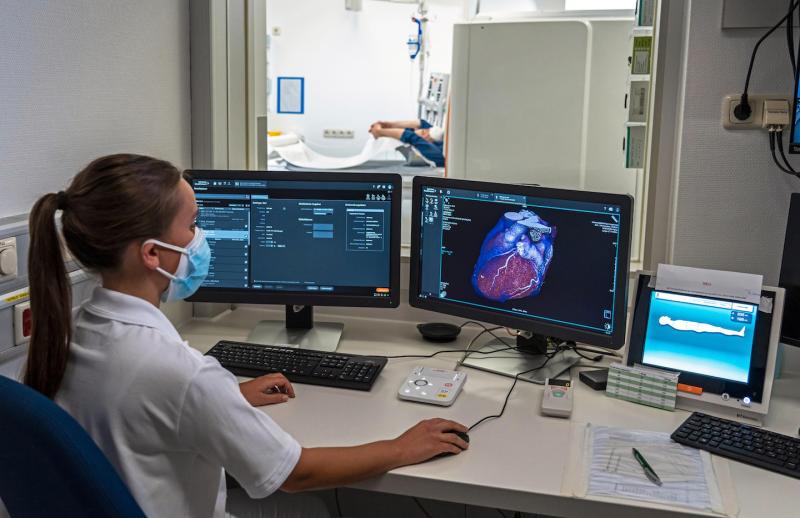
Feature | Computed Tomography (CT) | By Dave Fornell, DAIC Editor

The U.S. Food and Drug Administration (FDA) Sept. 30 cleared the world's first photon-counting computed tomography (CT)  ...

Home October 04, 2021
Home
Videos | Cardiovascular Information Systems (CVIS)

One of the trends in cardiovascular information system (CVIS) and radiology PACS at the Healthcare Information ...

Home August 31, 2021
Home
An example at HIMSS of deeper third-party software integration in enterprise imaging platforms was Siemens Healthcare showing a new, deep integration with echocardiography strain imaging analysis provider Epsilon Imaging. The integration eliminates the need for a separate login and the Epsilon software is now just a tool option available in the syngo system and it carries over data and images directly into the echo report.
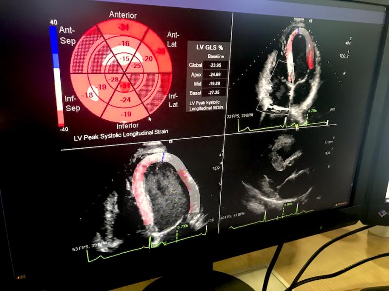
Feature | Information Technology | By Dave Fornell, Editor

Taking advantage of new technology advances, several radiology PACS, enterprise imaging and cardiovascular information ...

Home August 20, 2021
Home
Videos | Cardiovascular Ultrasound

A new ultrasound imaging technology that may offer novel ways to diagnose and better understand cardiac diseases using ...

Home August 17, 2021
Home
A comparison of color-flow Doppler cardiac ultrasound showing blood flow, and blood speckle tracking revealing a more detailed and complex understanding of the low. It shows the formation of a vortex that may play a role in future assessments for the efficiency of flow in the heart and vessels. #ASE21 #ASE2021 #Vectorflowimaging
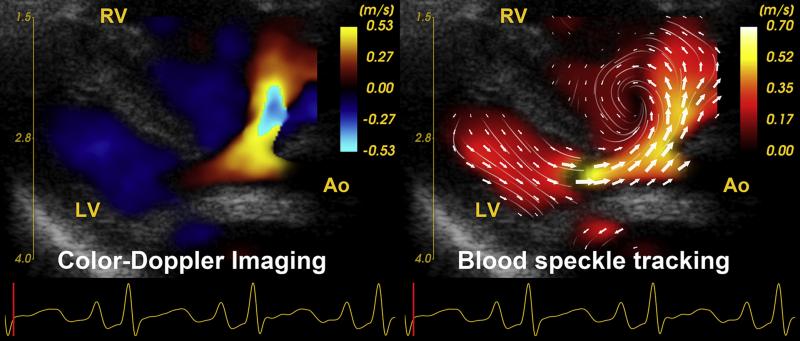
Feature | Cardiac Imaging | By Dave Fornell, Editor

In the past several years, a few cardiac ultrasound vendors have developed new ways to image the intricate flow of ...

Home August 17, 2021
Home
News | Medical 3-D Printing

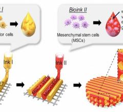
July 27, 2021 -- Cardiovascular diseases account for 32% of global deaths. Myocardial infarction, or heart attacks, play ...

Home July 27, 2021
Home
Images or a digital twin mitral valve of a patient created from cardiac ultrasound that were used to perform a virtual surgical procedure to test how the intervention would impact the patient prior to actually performing the procedure. The right image shows color coding for sheer stresses on the valve leaflets before and after the virtual surgery. The right image shows the model quantitation of leaflet coaptation at peak systole prior to the the virtual surgery. Read the original article in Plos One https:/
Feature | Cardiovascular Ultrasound | By Dave Fornell, Editor

Outside of medicine, computer-generated virtual twins of real machines like cars or airplanes have been used in ...

Home July 26, 2021
Home
News | Computed Tomography (CT)

July 16, 2021 — Canon Medical Systems USA is joining forces with Cleerly in a strategic partnership to support simple ...

Home July 16, 2021
Home
CentraCare completed the first structural heart procedure in the world guided by a new 4-D hologram technology developed by EchoPixel. The augmented reality technology aims to reduce reduce procedure time, improve accuracy of the procedure, and reduce risk of complications.
Feature | Virtual and Augmented Reality

June 28, 2021 — CentraCare, one of the largest health systems in Minnesota, successfully completed the first structural ...

Home June 28, 2021
Home
Illustration of how spectral CT can help confirm findings on a conventional CT scan without requiring follow-up imaging, such as a nuclear scan. The conventional CT on the left, and the dual-energy CT on the right confirms an infarct.
Feature | Computed Tomography (CT)

May 27, 2021 — Philips Healthcare released a workhorse computed tomography (CT) system, the Spectral CT 7500, which has ...

Home May 27, 2021
Home
Videos | Congenital Heart

Tom Jones, M.D., director, cardiac catheterization laboratories, Seattle Children’s Hospital, explains some of the new ...

Home May 26, 2021
Home
News | CT Angiography (CTA)

March 23, 2021 — Researchers at the Karolinska Institute in Stockholm, Sweden, have published the first-of-its-kind ...

Home March 23, 2021
Home
An example of 3-D computer aided design (CAD) software from Materialise being used at Henry Ford Hospital to evaluate left ventricular outflow tract (LVOT) obstruction from a Sapian TAVR valve virtually implanted in the mitral valve position. Henry Ford uses both 3-D printing and CAD to plan and guide complex structural heart procedures. 
Feature | Medical 3-D Printing | By Dave Fornell, Editor

With increasing complexity of interventional structural heart disease and congenital heart disease interventions, 3-D ...

Home January 24, 2021
Home
Subscribe Now