May 5, 2020 — The Society of Cardiovascular Computed Tomography (SCCT) announced plans this week to hold its annual ...
Computed Tomography (CT)
Cardiac computed tomography CT systems use a series of X-ray images to create an image volume dataset that can be sliced or manipulated on any plane using advanced visualization software. This channel includes content on CT scanners, CT contrast agents, CT angiography (CTA and CCTA), CT perfusion, spectral CT (also called dual souce or dual energy CT), and interative image reconstruction software that can reduce dose and make lower-quality CT images diagnostic.

 May 05, 2020
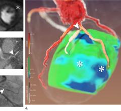
May 4, 2020 – A new technique that combines computed tomography (CT) and magnetic resonance imaging MRI can bolster ...
May 04, 2020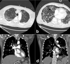
April 23, 2020 — A special report published in the journal Radiology outlines prevention, diagnosis and treatment of ...
April 30, 2020Sponsored Content

Blog | Enterprise Imaging
As medical advancements continue to push the boundaries of what is possible in the field of structural heart ...
August 10, 2023
Stephen Bloom, M.D., FASNC, director of nonivasive cardiology (cardiac CT, nuclear cardiology and echocardiography) at M ...
April 18, 2020
Feature | Coronavirus (COVID-19) | Dave Fornell, Editor
Cases of acute cardiovascular disease and cardiac complications caused by COVID-19 require cardiovascular imaging ...
April 18, 2020
Hicham Skali, M.D., a staff cardiologist and member of the Non-invasive Cardiovascular Imaging Program at Brigham and ...
April 04, 2020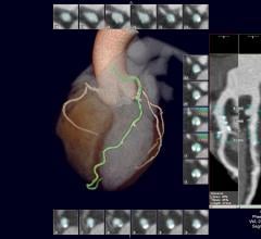
April 3, 2020 — Patients who experience chest pain and have abnormal results on a cardiac stress test but who do not ...
April 03, 2020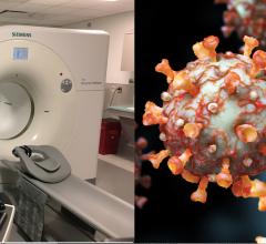
News | Coronavirus (COVID-19) | Dave Fornell, Editor
April 3, 2020 — A new guidance document on best practices to maintain safety and minimize contamination in nuclear ...
April 03, 2020
Feature | Interventional Radiology | By Katie Caron
A new year — and decade — offers the opportunity to reflect on the advancements and challenges of years gone by and ...
April 03, 2020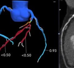
James Udelson, M.D., chief of the division of cardiology, Tufts Medical Center, explains how cardiac computed tomography ...
March 26, 2020
March 16, 2020 — The Society of Cardiovascular Computed Tomography (SCCT) released a new expert consensus document on ...
March 16, 2020
March 5, 2020 — Experts in the medical imaging community have developed a new, landmark consensus document to optimize ...
March 05, 2020
February 28, 2020 — New healthcare technologies are being implemented in the fight against the novel coronavirus (COVID ...
February 28, 2020
Feature | Artificial Intelligence | Sanjay Parekh, Ph.D.
February 7, 2020 – At the 2019 Radiological Society of North America (RSNA) meeting in December, there was a record ...
February 11, 2020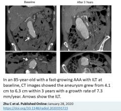
January 29, 2020 – The presence of a blood clot on the wall of the aorta in people with abdominal aortic aneurysms (AAA) ...
January 29, 2020© Copyright Wainscot Media. All Rights Reserved.
Subscribe Now