Videos

Cardiac Imaging

Coronavirus (COVID-19) | April 18, 2020

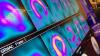
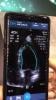
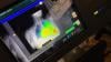
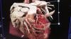
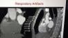
Stephen Bloom, M.D., FASNC, director of nonivasive cardiology (cardiac CT, nuclear cardiology and echocardiography) at Midwest Heart and Vascular Associates, Overland Park, Kansas. He is also a member of the American Society of Nuclear Cardiology (ASNC) Board of Directors, explains some of the issues involved and protocols used for cardiac imaging during the COVID-19 pandemic. His discussion includes computed tomography, cardiac ultrasound and nuclear imaging.

Right now, Bloom said it is difficult to test everybody and there is a shortage of masks, gowns and other personal protective equipment (PPE), and the imaging equipment needs to be sanitized each time it is used. He said it is just is not possible to image all the patients who need imaging right now. Hospitals also are trying to limit the number of healthy people people coming into hospitals for routine visits and tests to reduce their potential exposure to the novel coronavirus (COVID-19, SARS-CoV-2) and help containment efforts. 

"The tests should be done, very simply, if it changes the care of the patient. If it doesn't change the care of the patient, and it can be postponed, it should be postponed," Bloom explained. "I would say 80 percent of our cardiac imaging exams have stopped. It has been very dramatic."


Related Imaging Precautions During COVID-19 Content:

Cardiac Imaging Best Practices During the COVID-19 Pandemic

Best Practices for Nuclear Cardiology Laboratories During the Coronavirus (COVID-19) Pandemic

ASE Guidelines for the Protection of Echocardiography Providers During the COVID-19 Outbreak 

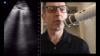
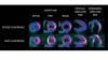
VIDEO: Best Practices for Nuclear Cardiology During the COVID-19 Pandemic — Interview with Hicham Skali, M.D.

VIDEO: Cancelling Non-essential Cardiac Procedures During the COVID-19 Outbreak — Interview with Ehtisham Mahmud, M.D. 

VIDEO: 9 Cardiologists Share COVID-19 Takeaways From Across the U.S.  

VIDEO: Telemedicine in Cardiology and Medical Imaging During COVID-19 — Interview with Regina Druz, M.D.

VIDEO: Use of Teleradiology During the COVID-19 Pandemic — an interview with radiologist John Kim, M.D.

Study Looks at CT Findings of COVID-19 Through Recovery

Experts Stress Radiology Preparedness for COVID-19

VIDEO: Imaging COVID-19 With Point-of-Care Ultrasound (POCUS) — Interview with emergency physician Mike Stone, M.D.,

VIDEO: How China Leveraged Health IT to Combat COVID-19 — Interview with Jilan Liu, M.D., CEO for the HIMSS Greater China

ACR Recommendations for the Use of Chest Radiography and CT for Suspected COVID-19 Cases

VIDEO: What Cardiologists Need to Know about COVID-19 — Interview with Thomas Maddox, M.D.

The Cardiac Implications of Novel Coronavirus


Coronavirus (COVID-19) | April 17, 2020

DAIC Editor Dave Fornell has conducted numerous video interviews remotely from his home office in March and April 2020 with nine cardiologists from around the United States. After each interview he asked how COVID-19 has impacted their hospital and them personally. This video offers a candid overview of their thoughts in the fight against the novel coronavirus.

Each was interviewed for other videos and some of the comments used here were from a questions after the main interview on how novel coronavirus (COVID-19, SARS-CoV-2) is impacting them, their patients or their cardiology departments. 

Insights include the following doctors. Click on the names to see their videos from March and April 2020:

   • Thomas Maddox, M.D., Washington University School of Medicine, St. Louis
   • Ehtisham Mahmud, M.D., UC San Diego Medical Center
   • William O’Neill, M.D., Henry Ford Hospital, Detroit
   • Regina Druz, M.D., Integrative Cardiology Center of Long Island, N.Y.
   • Justin Fried, M.D., Columbia University Irving Medical Center, New York City
   • Hicham Skali, M.D., Brigham and Women’s Hospital, Boston
   • Stephen Bloom, M.D., FASNC, Midwest Heart and Vascular Associates, Overland Park, Kansas
   • Michael Mack, M.D., Baylor Scott and White, Dallas, Texas
   • Basel Ramlawi, M.D, Heart and Vascular Center at Valley Health System in Virginia

Find more videos and news on the impact of COVID-19 on cardiology


Coronavirus (COVID-19) | April 04, 2020

Hicham Skali, M.D., a staff cardiologist and member of the Non-invasive Cardiovascular Imaging Program at Brigham and Women’s Hospital (BWH), and at Brigham and Women’s / Massachusetts General Health Care Center at Foxborough, explains the new recommendations from the American Society of Nuclear Cardiology (ASNC) and from imagers in China and Singapore. The ASNC created a best practices document for nuclear cardiology laboratories during the novel coronavirus (COVID-19, SARS-CoV-2) pandemic. The suggestions in the guidelines can ally to any imaging modality, including computed tomography (CT), MRI and ultrasound. 

Skali elaborates on the following points in his discussion, which are specific recommendations in the ASNC and SNMMI COVID-19 guidance document:
   • Rescheduling non-urgent visits
   • Rescheduling elective surgeries and procedures
   • Using separate spaces for patients with known or suspected COVID-19 to prevent spread
   • Ensuring supplies are available
   • Promoting use of telehealth
   • Screen staff, patients and visitors before they enter the department
   • Minimize non-essential visitors into the department
   • Record symptoms at the start of the shift
   • Use personal protective equipment (PPE)for healthcare personnel
   • If available, use PPE for patients due to concern of asymptomatic transmission of COVID-19
   • Maintain strict hand hygiene
   • Maintain 6 feet distance in all patient/staff interactions when possible
   • Work remotely whenever feasible, especially with ready studies
   • Rotating staff schedules for on-site and off-site work
   • Use of rest only studies if possible
   • Use of half-time SPECT to speed exam times
   • Use of PET if available to speed exam times

Skali served as the moderator in for the ASNC on demand webinar COVID-19 Preparedness for Nuclear Cardiology Labs: Insights from the US, China and Singapore.

VIDEO: Telemedicine in Cardiology and Medical Imaging During COVID-19 — Interview with Regina Druz, M.D., an ASNC Board member and also a speaker during the ASNC webinar.

Find more news and video on relating to COVID-19 and its impact on cardiology

FFR Technologies | March 26, 2020

James Udelson, M.D., chief of the division of cardiology, Tufts Medical Center, explains how cardiac computed tomography (CT) scans are being used to create image-derived fractional flow reserve (FFR) values to determine if a coronary lesion is flow limiting. The FFR-CT can help determine if the patient needs a stent, or if the disease can be treated with medication. Tufts uses FFR-CT evaluations on non-emergency chest pain patients to reduce the need for diagnostic catheterizations. 


Related FFR-CT Content:

Image-based FFR May Replace Pressure Wires and Adenosine

New Technology Directions in Fractional Flow Reserve (FFR)

8 Cardiovascular Technologies to Watch in 2020

VIDEO: Using FFR-CT in Everyday Practice

FFR-CT is Ready for Prime-time Evaluation of Coronary Disease

6 Hot Topics in Interventional Cardiology at TCT 2019

FFR-CT: Is It Radiology or Cardiology?


Find more news and video from Tufts Medical Center


Coronavirus (COVID-19) | March 20, 2020

An interview with Ehtisham Mahmud, M.D., FSCAI, chief, Division of Cardiovascular Medicine, executive director of medicine, Cardiovascular Institute, director of  interventional cardiology and cardiac cath lab at UC San Diego Medical Center, and president of the Society for Cardiovascular Angiography and Interventions (SCAI). He explains the how cardiology departments in the U.S. are now postponing cardiovascular procedures due to novel coronavirus (COVID-19, SARS-CoV-2) containment efforts and new guidelines from Medicare calling for delay of all elective procedures in the country. 

Mahmud explains how patients are being prioritized, with acute myocardial infarction patients or others with acute, life-threatening conditions, or at high risk for a near term hospital admission, will still receive cardiac catheterizations, cardiovascular surgery or structural heart procedures for MitraClip and transcatheter aortic replacement (TAVR) under certain circumstances. All other procedures are being postponed until further notice based in the spread and infection rates of COVID-19. He said most hospitals, including his own, are moving to telehealth visits via phone or online to continue clinic work with patients, including those with chronic conditions such as heart failure.

The Centers for Medicare and Medicaid Services (CMS) announced March 18, 2020, that all elective surgeries, and non-essential medical, surgical and dental procedures should be delayed during the coronavirus outbreak. This move is three-fold. 
   1. It is to help with containment efforts by reducing patient and family travel to hospitals, which are at the center of the COVID-19 outbreak. 
   2. Delaying procedures will help preserve and inventory of personal protective equipment (PPE), hospital beds and ventilators and other medical supplies. 
   3. With the start of social distancing and the shut down of all large gatherings, this has severely impacted blood drives and other blood donations, so the nation's blood banks have severely limited supplies.

“The reality is clear and the stakes are high — we need to preserve personal protective equipment for those on the front lines of this fight,” said CMS Administrator Seema Verma.

This will not only preserve equipment but also free up the healthcare workforce to care for the patients who are most in need. Additionally, as states and the nation as a whole work toward limiting the spread of COVID-19, healthcare providers should encourage patients to remain home, unless there is an emergency, to protect others while also limiting their exposure to the virus. 

Read Mahmud's SCAI President's letter The Evolving Pandemic of COVID-19 and Interventional Cardiology


Related Cardiology Related COVID-19 Content:

ACC COVID-19 recommendations for the cardiovascular care team

VIDEO: What Cardiologists Need to Know about COVID-19 — Interview with Thomas Maddox, M.D.

The Cardiac Implications of Novel Coronavirus

ESC Council on Hypertension Says ACE-I and ARBs Do Not Increase COVID-19 Mortality

VIDEO: Imaging COVID-19 With Point-of-Care Ultrasound (POCUS)

CT Provides Best Diagnosis for Novel Coronavirus (COVID-19)

Radiology Lessons for Coronavirus From the SARS and MERS Epidemics

Deployment of Health IT in China’s Fight Against the COVID-19 Epidemic

Emerging Technologies Proving Value in Chinese Coronavirus Fight

Radiologists Describe Coronavirus CT Imaging Features

Coronavirus Update from the FDA

CT Imaging of the 2019 Novel Coronavirus (2019-nCoV) Pneumonia

CT Imaging Features of 2019 Novel Coronavirus (2019-nCoV)

Chest CT Findings of Patients Infected With Novel Coronavirus 2019-nCoV Pneumonia 


Additional COVID-19 Resources for Clinicians:

   ACC COVID-19 Hub page   

   Johns Hopkins Coronavirus Resource Center with inteavtive map of cases in U.S. and worldwide 

   World Health Organization (WHO) COVID-19 situation reports

   World Health Organization (WHO) coronavirus information page

   U.S. Food and Drug Administration (FDA) COVID-19 information page

   Centers for Disease Control (CDC) COVID-19 information page

   Centers for Medicare and Medicaid Services (CMS) frequently asked questions and answers (FAQs) for healthcare providers regarding COVID-19 related payments


Coronavirus (COVID-19) | March 17, 2020

Interview with Mike Stone, M.D., an emergency physician at Northwest Acute Care Specialists in Portland, Ore., director of point of care ultrasound education at Butterfly Network Inc., and and former chief of the division of emergency ultrasound at Brigham and Women's Hospital, Boston. He explains what clinicians look for in lung ultrasound to evaluate patients for plural wall thickening and areas of congestion inside the lung typical of pneumonia and seen in patients with novel coronavirus (COVID-19, also referred to clinically as SARS-COV-2). 

Small hand-held ultrasound systems may offer advantages over X-ray or computed tomography (CT), because the entire system can be contained inside a protective sheath, making it easier to disinfect the device rather than an entire imaging room. Point of care ultrasound (POCUS) systems also allow triage of patients in tents or other areas outside of the hospital where other imaging modalities are not available. 

Stone shares examples of COVID-19 ultrasound images from Butterfly Network POCUS system technology, which turns an iPhone into an FDA-cleared ultrasound device for multiple POCUS exam types. It is the first POCUS system reported to be used for triage and monitoring COVID-19 patients. 
 

Related Content:

VIDEO: Point of Care Ultrasound (POCUS) in Cardiology and Critical Care — Interview with Michael Lanspa, M.D.

VIDEO: Automated Cardiac Ejection Fraction for Point-of-care-ultrasound Using Artificial Intelligence

The Cardiac Implications of Novel Coronavirus

VIDEO: What Cardiologists Need to Know about COVID-19 — Interview with Thomas Maddox, M.D.

ESC Council on Hypertension Says ACE-I and ARBs Do Not Increase COVID-19 Mortality

Find more related clinical content Coronavirus (COVID-19)

Study Looks at CT Findings of COVID-19 Through Recovery

Experts Stress Radiology Preparedness for COVID-19

ACR Recommendations for the Use of Chest Radiography and CT for Suspected COVID-19 Cases

Wuhan CT Scans Reliable for Coronavirus Diagnosis, Limited for Differentiation

RSNA | January 13, 2020

DAIC/ITN Editor Dave Fornell takes a tour of some of the most innovative new medical imaging technologies displayed on the expo floor at the Radiological Society of North America (RSNA) 2019 meeting. 

Technology examples include a robotic arm to perform remote ultrasound exams, integration of artificial intelligence (AI) to speed or automate radiology workflow, holographic medical imaging display screens, a new glassless digital radiography (DR) X-ray detector, augmented reality for transesophageal echo (TEE) training, moving DR X-ray images, 3-D printed surgical implants created from a patient's CT imaging, DR X-ray tomosynthesis datasets, radiation dose management and analytics software, and new computed tomography (CT) technologies.


Find more videos and news from RSNA 2019


Magnetic Resonance Imaging (MRI) | January 06, 2020

Karen Ordovas, M.D., MAS, professor of radiology and cardiology at the University of California San Francisco (UCFS) School of Medicine and a Society of Cardiac Magnetic Resonance (SCMR) board member, explains how cardiac MRI can help in women's heart disease and to better define complex congenital heart anatomy. She spoke at the 2019 Radiological Society of North America (RSNA) meeting. 

She specializes in cardiac and pulmonary imaging, and has particular expertise in using CT and MRI techniques in cardiovascular imaging and the differences of presentation in imaging between male and female cardiac patients. Ordovas is helping advance education around heart disease in women and bring great awareness of quality tools to diagnose heart disease and how heart MRI can help. She also is heavily involved in the use of heart MRIs for pregnant women, since there is no radiation,  and patients with congenital heart disease where detailed imaging of the complex anatomy is required.

The use of cardiac MRI in congenital heart disease is common in serial imaging of patients with Tetralogy of Fallot (TOF), one of the most common congenital heart diseases for which patients are referred for post-operative magnetic resonance (MR) imaging evaluation. In the past few decades, surgery has proved successful, but most patients require repeat imaging throughout their lives and MRI can offer more detailed soft tissue imaging without the use of radiation. 


Related Cardiac MRI Content:

Cardiac MRI Becoming More Widely Available Thanks to AI and Reduced Exam Times

VIDEO: Advances in Cardiac MRI Technology — Interview with James Carr, M.D.

Cardiac MRI Delivers Accurate Diagnosis for Frontline Chest Pain Evaluation

VIDEO: Dedicated Cardiac MRI Use at the Baylor Scott White Heart Hospital

Advantages and New Applications of Cardiac MRI

Will Cardiac MRI Expand?


Magnetic Resonance Imaging (MRI) | December 20, 2019

James Carr, M.D., chair of the Department of Radiology, Northwestern University, and incoming 2020 President of the Society of Cardiac Magnetic Resonance (SCMR), explains why MRI is an ideal cardiac imaging modality, at the 2019 Radiological Society of North America (RSNA) meeting.

Heart MRI offers advantages over computed tomography (CT) and echocardiography because of its excellent soft tissue delineation and its ability to offer information beyond anatomical imaging, such as perfusion, morphology and metabolism. MRI can be technically challenging and the exams requiring a long time, but recent advances have helped cur cardiac imaging times down significantly. Automation and artificial intelligence (AI) also is making post-processing and quantification mush faster, brining it closer to the time it takes to scan and post-process CT imaging.

Northwestern is was one of the early adopters of cardiovascular MRI. Carr said heart MRI was not common in regular clinical use until the past decade at some luminary centers. In 2005, Carr was given the opportunity to develop a clinical cardiac MRI program at Northwestern.
 
He said MRI scanners have improved, and now much faster than a decade ago. They are also more optimized for cardiac imaging. While heart MRI is well known in large hospital centers, Carr said it still needs to develop and expand to community hospitals and rural hospitals outside major population centers. 

Artificial intelligence is playing a significant role in cardiac MRI automation of speeding workflow and quantification. Carr said these technologies will become mainstream in the next few years. AI also will play an increasing role in risk prediction based on new image analysis algorithms in development.

For more information on cardiac MRI, visit SCMR's website www.heartmri.org.


Related Cardiac MRI Content:

Cardiac MRI Delivers Accurate Diagnosis for Frontline Chest Pain Evaluation

Cardiac MRI Becoming More Widely Available Thanks to AI and Reduced Exam Times

VIDEO: Dedicated Cardiac MRI Use at the Baylor Scott White Heart Hospital — Interview with Haojie Wang, M.D.

VIDEO: Dedicated Cardiac MRI Use at the Baylor Scott White Heart Hospital

Advantages and New Applications of Cardiac MRI

Will Cardiac MRI Expand?

VIDEO: Use of Cardiac MRI in Congenital and Women's Heart Disease — Karen Ordovas, M.D., 


Cardiovascular Ultrasound | December 20, 2019

This is the LVivo auto cardiac ejection fraction (EF) app that uses artificial intelligence (AI) from the vendor Dia, displayed at the Radiological Society Of North America (RSNA) 2019. The user opens the app in a couple seconds the AI defines to myocardial border and calculates EF for left ventricle (LV). It is shown here integrated into the GE Healthcare VScan point-of-care-ultrasound system (POCUS).

The company also partners with Konica-Minolta to supply auto EF for ultrasound images on its cardio PACS.

Read more about the system from an ASE 2019 stud

Radiation Dose Management | December 19, 2019


Mahadevappa Mahesh, Ph.D., chief of medical physicist and professor of radiology and medical physics, Johns Hopkins University, Baltimore, treasurer of the American Association of Physicists in Medicine (AAPM),a board member of the American College of Radiology (ACR), presented a late-breaking study on how medical imaging radiation dose has started to drop over the past decade. He is the co-chair of the National Council on Radiation Protection and Measures Report (NCRP), and presented the most recent NCRP data analysis at the 2019 Radiological Society of North America (RSNA) meeting.

The new NCRP 184 report covers the period between 2006 and 2016, the period of the most current CMS data. It shows a decrease of about 20 percent in the radiation dose the U.S. population receives from medical imaging, compared to the NCRP 160 that covered the period of up to 2006.

Key findings of the study include:

   • CT dose dropped about 6 percent, despite a 20 percent increase CT scans since 2006;

   • Drop of more than 50 percent for nuclear imaging scans, mainly due to fewer procedures begin performed;

   • A 15-20 percent decrease across X-ray imaging modalities.

Mahesh says this shows the impact of using "as low as reasonably achievable" (ALARA) principals, new dose guidelines outlined jointly by numerous medical societies, and dose reduction initiatives like Image Wisely, Image Gently, and the American College of radiology (ACR) Dose Index Registry.

He said there was growing concern a decade ago when the last council report was published, which show a steep increase in radiation dose. This was mainly due to a rapid increase in the use of computed tomography (CT) and other types of X-ray based and nuclear radiotracer medical imaging. This prompted the ACR top create the Image Wisely program and push for the use of more thoughtful imaging doses based on patient size, using the "as low as reasonably achievable” (ALARA) principle. While CT dose was lowered, he said the biggest decline was in nuclear imaging.


Cardiovascular Ultrasound | December 19, 2019

This is an example of an augmented reality (AR) training system for transesophageal echo (TEE) created by the simulation company CAE. Rather than just looking at an overhead screen, this system allows the user to use a HoloLens visor to see the impact their probe manipulation has on the imaging and better shows the orientation of the ultrasound probe, the 2-D ultrasound image slice and the relation to the anatomy. It was displayed at the 2019 Radiological Society Of North America (RSNA) meeting.

Read more about this technology.

Find more technology news and video from the RSNA meeting


RSNA | December 18, 2019

DAIC Editor Dave Fornell and Imaging Technology News (ITN) Consulting Editor Greg Freiherr offer a post-game report on the trends and technologies they saw on the expo floor of 2019 Radiological Society of North America (RSNA) annual meeting. This includes artificial intelligence (AI), augmented reality, holographic imaging, cybersecurity, and advances in digital radiography (DR) with a glassless detector plate, X-ray tomosynthesis, dual-energy X-ray and dynamic DR imaging. 

Find RSNA news and other videos

Nuclear Imaging | November 07, 2019

Rob Beanlands, M.D., FASNC, 2019 American Society of Nuclear Cardiology (ASNC) president, shares a couple trends he sees in cardiac nuclear imaging. He is the Vered Chair and division head of cardiology and director of the National Cardiac PET Centre at the University of Ottawa Heart Institute, Canada.

He said overall trends he sees in nuclear cardiology include the use of better myocardial reserve quantification so it is clear whether revascularization would help patients. Beanlands also said there is increasing interest in positron emission tomography (PET) imaging because of its superior image quality and increasing access to PET radiotracers. New tracers on the horizon will also increase the image quality and flexibility of PET to accommodate exercise stress.

He spoke on artificial intelligence (AI) development for medical imaging in sessions at the 2019 American Society of Nuclear Cardiology (ASNC) annual meeting. 

Artificial Intelligence | November 07, 2019

Piotr J. Slomka, Ph.D., FACC, research scientist in the Artificial Intelligence in Medicine Program, Department of Medicine at Cedars-Sinai, and professor of medicine in-residence of the David Geffen School of Medicine, UCLA. He explains how his team at Cedars-Sinai is working on intelligent patient risk prediction algorithms that will automatically extract information from medical imaging. He spoke on artificial intelligence (AI) development for medical imaging in sessions at the 2019 American Society of Nuclear Cardiology (ASNC) annual meeting. 

Find more articles and video on AI


Nuclear Imaging | November 06, 2019

Robert C. Hendel, M.D., explains some of the new cardiac radiotracers in the pipeline that were discussed in sessions at the American Society of Nuclear Cardiology (ASNC) 2019 meeting. Hendel is a professor of medicine and radiology and the Sidney W. And Marilyn S. Lassen Chair in Cardiovascular Medicine, Tulane University. He also serves as the chief of the section of cardiology and director of the Tulane University Heart and Vascular Institute. 

He outlined three new radiotracers that may impact cardiac imaging:

   • Flurpiridaz F-18 PET perfusion agent that offers high quality images and can be used with exercise stress;

   • MIBG imaging to help better assess heart failure classes and if patients may need an ICD;

   • New F-18 agents to image cardiac amyloidosis; and

   • Nuclear agents to enable annexin imaging, allowing direct vsualization of myocardial apoptotic cells.


PET-CT | October 30, 2019

Rupa Sanghani, M.D., FASNC, associate professor, Rush Medical College, director of nuclear cardiology and stress laboratory, Rush University Medical Center, and associate director, Rush Heart Center for Women, explains how to create a high-volume cardiac positron emission tomography (PET) imaging program. She spoke on this topic at the 2019 meeting of the American Society Nuclear Cardiology (ASNC) and led a tour for attendees of the PET-CT system at Rush, which was located close to the conference. 

Sanghani said the advantages of PET myocardial perfusion imaging include faster exam times and allowing additional information from coronary reserve flow assessments to better understand if revascularization will help a patient's heart recover. The 16-slice CT scanner is used not only to attenuate the PET images, but to perform a CT calcium scoring exam to assess the patient's risk for future cardiovascular events. The Rubidium-82 radiotracer used for PET exams only has a 75 second half life, so it can help increase the number of exams a center is able to perform each day. At higher volume centers, PET is optimized to handle all the patients who require pharmacological stress exams. 

In the video, Sanghani  outlines what Rush did to design its room, covers basics on training, what to look for in a scanner and other considerations when creating a PET program.

Read the article How to Start a Cardiac PET-CT Imaging Program, which includes more information from the Rush PET program.
 

Find more coverage of the ASNC

Find more news on nuclear imaging


Cardiac Diagnostics | October 29, 2019

Clyde Yancy, M.D., MSc, cardiology chief and vice dean for diversity and inclusion at Northwestern University, Feinberg School of Medicine, was a keynote speaker at the 2019 American Society of Nuclear Cardiology (ASNC) annual meeting. He said the traditional biases of seeing a patient and automatically making clinical assumptions because they are a certain race or gender are obsolete. For example, he said not all black patients have hypertension. Yancy added that genetics, especially with racial intermarriage over the past several generations, no longer predisposes patients to what is typically assumed for certain ethnic or racial backgrounds.

Yancy also said new research is showing how diet plays a major role in patient health and disease progression for things previously thought to be based on genetics. This includes the people who live in "food deserts" in urban areas where there are no sources of fresh food and vegetables, so they consume large amounts of packaged and processed foods that contain high levels of salt, phosphates and preservatives. He said these chemicals and diet may be the root cause of hypertension and diabetes in black populations in low income areas, rather than genetics as previously thought.


Related Content: 

VIDEO: Reducing Hypertension Among African-Americans — Interview with Kim Allan Williams, Sr., M.D.

VIDEO: Use of Plant-Based Diet to Reduce Cardiovascular Disease Risk — Interview with Kim Allan Williams, Sr., M.D.

VIDEO: New PLATINUM Diversity Data Shows Early DAPT Cessation OK in Minorities With New Generation Stent — Interview with Roxana Mehran, M.D., 


Structural Heart | October 16, 2019

Vivian Ng, M.D., assistant professor of medicine and an interventional cardiologist at the NewYork-Presbyterian/Columbia University Medical Center Structural Heart and Valve Center, helped organize the first Women in Structural Heart (WISH) event at the 2019 Transcatheter Cardiovascular Therapeutics (TCT) meeting. The evening session was standing room only and highlighted structural heart case presentations and discussion panels made up of all women. The session panelists and presenters were a whose-who of well known women in cardiology. The event was organized as a way to break the glass ceiling in the subspecialty of interventional cardiology, where women make up less than 5 percent of the operators.


Previous Video Interviews With Speakers and Panelists Involved in the WISH Session:

VIDEO: The Importance of the Neo-LVOT in Transcatheter Mitral Valve Replacement — Interview with Dee Dee Wang, M.D.

VIDEO: The Value of the Cardiovascular Service Line — Interview with Linda Gillam, M.D.,

VIDEO: Can We Live in 3-D Echo? — Interview with Lissa Sugeng, M.D.

VIDEO: Tricuspid Valve Imaging and Interventions Developing Hand-in-hand — Interview with Rebecca Hahn, M.D.

VIDEO: Strategies to Avoid Acute Kidney Injury Caused by Cath Lab Contrast — Interview with Roxana Mehran, M.D.,


Find more news and videos from TCT 2019

University of Colorado Hospital | October 02, 2019

Interview with John Carroll, M.D., director of interventional cardiology, Robert Quaife, M.D., director of advanced cardiac imaging, and James Chen, Ph.D., associate professor of medicine and director of the 3-D imaging lab at the Cardiac and Vascular Center at the University of Colorado Hospital. They discuss how the structural heart program was created and how they invested in advanced imaging to grow into one of the most advanced programs in the country. They explain how the program now incorporates transcatheter aortic valve replacement (TAVR), transcatheter mitral valve repair, transcatheter mitral valve replacement (TMVR), left atrial appendage (LAA) occlusion and transcatheter closure of holes in the heart. 

The heart team in this video stressed the need for advanced imaging to plan and guide the procedures. They explain how the center developed its own 3-D imaging software and worked with Philips healthcare to commercialize some of the technologies, including the EchoNavigator system used to fuse live angiography with live transesophageal echo (TEE).


Related University of Colorado Hospital Content:

Highlighting Innovation at the University of Colorado Hospital Cardiology Program

VIDEO: Evolution of Transcatheter Mitral Valve Repair at the University of Colorado — Interview with John Carroll, M.D., and Robert Quaife, M.D.

VIDEO: The Role of Advanced Imaging in Structural Heart Interventions — Interview with Robert Quaife, M.D.

VIDEO: Advice For Hospitals Starting a Structural Heart Program — Interview with John Carroll, M.D.

VIDEO: The Evolution of Complex PCI at University of Colorado — Interview with John Messenger, M.D., and Kevin Rogers, M.D.

VIDEO: Developing New Cath Lab Technologies With Real-time Collaboration Between Industry, Doctors

360 View of the TEE Echo Workstation During a MitraClip Procedure

VIDEO: Walk Through of a Hybrid Cath Lab at the University of Colorado Hospital

VIDEO: Cath Lab Walk Through at the University of Colorado Hospital

VIDEO: The Cardiac Surgeon Perspective on Transcatheter Mitral Valve Repair — Interview with Joe Cleveland, M.D.

VIDEO: An Overview of PFO Closure to Treat Cryptogenic Stroke — Interview with Karen Orjuela, M.D.,


(This video was originally posted in May 2019 and was updated Oct. 2, 2019)


Cath Lab | September 28, 2019

A discussion with Nicolas Bevins, Ph.D., vice chair, physics and research, and Jessica Harrington, RCIS. They explain the use of shields, technique and use of newer angiography technologies to reduce X-ray radiation dose in the cardiac cath labs at Henry Ford Hospital, Detroit.

Watch the VIDEO: Technologies and Techniques to Reduce Radiation Dose in the Cardiac Cath Lab — Interview with Akshay Khandelwal, M.D., director of medical operations at the Henry Ford Heart and Vascular Institute

Additional articles and videos on Henry Ford Hospital 


For more on how to reduce dose in the cath lan, read these related articles:

Cardiology Societies Call for Better Radiation Dose Tracking

Defining the Cath Lab Workplace Radiation Safety Hazard

Dose-Lowering Practices for Cath Lab Angiography

5 Technologies to Reduce Cath Lab Radiation Exposure

VIDEO: Heart Surgeon Shares Effects of Fluoroscopic Radiation Exposure

Helping Interventional Cardiologists Reduce Exposure to Ionizing Radiation

14 Ways to Reduce Radiation Exposure in the Cath Lab


(Editor's note - this video was originally published in September 2018 and was revised September 2019)

September 27, 2019

Akshay Khandelwal, M.D., director of medical operations at the Henry Ford Heart and Vascular Institute, Detroit, and associate professor of cardiology at Wayne State University, explains how his center has reduced X-ray radiation dose in the cardiac catheterization labs.

Watch the related VIDEO: Reducing Cath Lab Radiation Dose at Henry Ford Hospital — Discussion with Nicolas Bevins, Ph.D., vice chair, physics and research, and Jessica Harrington, RCIS, Henry Ford Hospital.

Additional articles and videos on Henry Ford Hospital 


For more on how to reduce dose in the cath lan, read these related articles:

Cardiology Societies Call for Better Radiation Dose Tracking

Defining the Cath Lab Workplace Radiation Safety Hazard

Dose-Lowering Practices for Cath Lab Angiography

5 Technologies to Reduce Cath Lab Radiation Exposure

VIDEO: Heart Surgeon Shares Effects of Fluoroscopic Radiation Exposure

Helping Interventional Cardiologists Reduce Exposure to Ionizing Radiation

14 Ways to Reduce Radiation Exposure in the Cath Lab


(Editor's note - this video was originally published in November 2018 and was revised September 2019)


Interventional Radiology | September 10, 2019

Scott Schwartz, M.D., interventional radiologist and program director for IR residencies and the vascular and interventional radiology fellowship at Henry Ford Hospital, explains how the department uses Yttrium-90 (Y90) embolization therapy to treat liver cancer.

Additional articles and videos on Henry Ford Hospital 


Related Articles on Y-90 Radiotherapy:

Current Advances in Targeted Radionuclide Therapy

A Look Ahead in Targeted Radionuclide Therapy

Radioactive Bead Therapy Now Used for Head, Neck Tumors

NCCN Guidelines Recommend Y-90 Microspheres for Metastatic Colorectal Cancer Treatment


(Editor's note - this video was originally published in November 2018 and was revised September 2019)

Advanced Visualization | August 09, 2019

An example of Siemens' photo-realistic Cinematic image reconstruction. This image is from a CTA exam from a Siemens Force CT scanner. 

Vendors who offer this realistic type of CT image rendering say it is not used for diagnostics. However, the technology can be helpful when explaining things to the patient and their family, educating physicians and staff, and for surgeons, since it offers a realistic view of the anatomy that is easier for most people to understand who are not familiar with cardiac anatomy as it appears in traditional CT multiplanar reconstruction (MPR) images. 

This example of software was demonstrated on the expo floor at the 2019 Society of Cardiovascular Computed Tomography (SCCT) meeting. 


Find more news and videos from the SCCT meeting

CT Angiography (CTA) | August 08, 2019

This is a quick video example of a cardiac computed tomography (CT) exam showing a Medtronic CoreValve transcatheter aortic valve replacement (TAVR) device implanted. The image was reconstructed using Canon Medical’s Global Illumination photo-realistic rendering advanced visualization post-processing software. Vendors who offer this realistic type of CT image rendering say it is not used for diagnostics. However, the technology can be helpful when explaining things to the patient and their family, educating physicians and staff, and for surgeons, since it offers a realistic view of the anatomy that is easier for most people to understand who are not familiar with cardiac anatomy as it appears in traditional CT multiplanar reconstruction (MPR) images. 

This example of software was demonstrated on the expo floor at the 2019 Society of Cardiovascular Computed Tomography (SCCT) meeting. 


CT Angiography (CTA) | August 08, 2019

This is an example of an automated calcium scoring software to speed review of coronary artery calcium (CAC) scoring cardiac computed tomography (CT) scans. This advanced visualization software from Ziosoft uses artificial intelligence to segment the coronary vessels, identify valves and the aorta and then color code tag the calcium deposits and quantify the amount of calcified plaque in each vessel. It tallies the score into a table and computes an overall Agatston risk score. This risk score correlates to that patient's risk for a heart attack in the future. The software notes calcium in the heart outside the coronaries in valve leaflets and the aorta, but excludes this data. This type of automation is now offered by most advanced visualization and CT system vendors. This automation can save a large amount of post-processing time and make it easier for hospitals to offer low-cost CAC CT screening programs. 

CAC scans can be used to determine if a patient needs to go on statin therapy. An Agatston score of zero means the patient has no risk of coronary disease. 

Calcium in arteries is a marker for damage caused by vessel wall inflammation from atherosclerosis. Calcium can form from previously ruptured necrotic, lipid core plaques, also referred to as vulnerable plaques. These are the types of plaque responsible for heart attacks. When the core of these plaques rupture, the blood reacts to the exposed core similar to a wound and begins to clot, forming a thrombus in the vessel, which can block the blood flow. When the vessel heals over time it calcifies, leaving behind an easily identifiable marker on CT imaging. 

This example of software was demonstrated on the expo floor at the 2019 Society of Cardiovascular Computed Tomography (SCCT) meeting. 


Related CT Calcium Scorining Content:

VIDEO: The History of CT Calcium Scoring — Interview with Arthur Agatston, M.D.

VIDEO: New Cholesterol Guidelines Support CT Calcium Scoring for Risk Assessment — Interview with Matthew Budoff, M.D.

CT Calcium Scoring Becoming a Key Risk Factor Assessment

ACC and AHA Release Updated Cholesterol Guidelines for 2018

VIDEO: CT Calcium Scoring to Screen For Who Should Take Statins — Interview with Matthew Budoff, M.D.


Computed Tomography (CT) | August 07, 2019

This is an example of in-stent restenosis shown using spectral CT imaging. This example was demonstrated by Philips Healthcare at the 2019 meeting of the Society Of Cardiovascular Computed Tomography (SCCT). It can sometimes be difficult to determine if showing inside a stent of CT is artifact or real. Spectral CT can definitely help make a diagnosis. This example shows Philips’ Effective Z visualization, where the image is created based on the atomic number of the elements present in the image. After removal of iodine from the image, it shows the shadow in the stent has the same atomic composition as the vessel intima.  

This example is from a Philips IQon dual-energy CT scanner. This system has spectral imaging always on without any need to change protocols. This allows the spectral to be used when needed for additional information without need to rescan a patient.

Read more on this CT system.

VIDEO: Applications of Spectral CT — Interview with Suhny Abbara, M.D.


Cardiac Imaging | July 30, 2019

Nate Bachman, graduate research assistant in the Human Cardiovascular Physiology Lab of the Dept. of Health and Exercise Science at Colorado State University, describes how he and fellow researchers used multiple types of cardiac imaging to evaluate the cardiovascular health of athletes who compete in endurance events lasting six hours or more, and what the results may suggest for future screening.

EP Lab | July 26, 2019

Mark Ibrahim, M.D., FACC, assistant professor of medicine and radiology, associate program director, advanced cardiac imaging fellowship, University of Utah, explains what radiologists and cardiologists need to know what is needed from CT imaging prior to ablation procedures for atrial fibrillation (AF) and ventricular fibrillation (VF). He spoke at a joint session of the Heart Rhythm Society (HRS) and the Society of Cardiovascular Computed Tomography (SCCT) at the 2019 SCCT meeting. 


Find more SCCT news and videos

CT Angiography (CTA) | July 26, 2019

Andrew Choi, M.D., FACC, FSCCT, co-director, cardiac CT and MRI, assistant professor of medicine and radiology, George Washington University, Division of Cardiology, Washington, D.C., explains the role of social media to extend the reach of cardiovascular research from peer review journals. He spoke on this topic in multiple sessions at the Society of Cardiovascular Computed Tomography (SCCT) 2019 meeting.


Find more SCCT news and videos


EP Lab | July 25, 2019
Heart Valve Technology | July 24, 2019

Joao Cavalcante, M.D., FSCCT, director of structural heart CT and cardiac MRI, Minneapolis Heart Institute, discusses transcatheter mitral valve replacement (TMVR) imaging requirements at the Society of Cardiovascular Computed Tomography (SCCT) 2019 meeting. SCCT held its first TMVR planning hands-on workshop, which he was involved in. TMVR is expanding with currently FDA cleared valve-in-valve (VIV) and valve-in-ring (VIR) procedures.


Related TMVR Content:

Interventional Imagers: The Conductors of the Heart Team Orchestra

VIDEO: The Importance of the Neo-LVOT in Transcatheter Mitral Valve Replacement  — Interview with Dee Dee Wang, M.D.

VIDEO: Transcatheter Structural Heart Procedure Navigation Technology Advances  — Interview with Stephen Little, M.D.

Recent Advances in Transcatheter Valve Technology

Abbott Begins Tendyne Transcatheter Mitral Valve U.S. Pivotal Trial

VIDEO: The Essentials of CT Transcatheter Valve Imaging — Interview with Jonathon Leipsic, M.D.,

New LAMPOON Technique Reduces LVOT Obstruction in Transcatheter Mitral Valve Replacement

Find more SCCT news and videos

CT Angiography (CTA) | July 24, 2019

Arthur Agatston, M.D., clinical professor of medicine, Florida International University, Herbert Wertheim College of Medicine, is the name-sake of the Agatston score used in CT calcium scoring. He explains the history of the scoring system from the early 1990s and the evolution of CT technology for cardiac imaging. The latest American Heart Association (AHA) 2018 cholesterol guidelines now include the use of CT calcium scoring, which was a big topic at the Society of Cardiovascular Computed Tomography (SCCT) 2019 meeting.

Read the article How the Agatston Calcium Score Was Created and its Impact on Heart Attack Prevention.

See a quick example of a CT calcium scoring exam in the VIDEO: Example of an Automated CT Cardiac Calcium Scoring Exam.


Related CT Calcium Scorining Content:

VIDEO: New Cholesterol Guidelines Support CT Calcium Scoring for Risk Assessment — Interview with Matthew Budoff, M.D.

CT Calcium Scoring Becoming a Key Risk Factor Assessment

ACC and AHA Release Updated Cholesterol Guidelines for 2018

VIDEO: CT Calcium Scoring to Screen For Who Should Take Statins — Interview with Matthew Budoff, M.D.


Find more SCCT news and videos


SCCT | July 19, 2019

Ron Blankstein, M.D., director of cardiac computed tomography, Brigham and Women's Hospital, and associate professor of medicine and radiology, Harvard Medical School, and current SCCT president, offers an overview of the recent trends in cardiac CT and some of the new highlights at the Society of Cardiovascular Computed Tomography (SCCT) 2019 meeting. The  said key topics included integration of artificial intelligence into CT systems, the integration of CT calcium scoring into the 2018 American Heart Association (AHA) cholesterol management guidelines, structural heart assessments for transcatheter valve and left atrial appendage (LAA) occlusion, and partner sessions with TCT and HRS that explain the roll of CT in interventional cardiology and electrophysiology.

Find more SCCT news and videos

CT Angiography (CTA) | July 19, 2019

Quynh Truong, M.D., MPH, associate professor of radiology and medicine at Weill Cornell and director of cardiac CT, NewYork Presbyterian Hospital, offers 10 tips to help improve image quality for cardiovascular computed tomography (CTA) exams. She spoke on this topic at the Society of Cardiovascular Computed Tomography (SCCT) 2019 meeting.

Her discussion includes:

1. Practice the breath hold

2. Give nitrate to vasodilate

3. Slow the heart rate

4. Acquire multiple phases with padding

5. Include systolic imaging for irregular heart beats

6. Consider cauo-cranial imaging to avoid contrast enhancement issues

7. Use a high contrast injection rate

8. Right ventricular opacification for function

9. Use a hard kernel

10. Know your post-processing software


VIDEO: Tips and Tricks to Aid Cardiac CT Technologist Workflow

VIDEO: How to Prepare a Patient for a Cardiac CT Scan

Find more SCCT news and videos

Cardiovascular Ultrasound | July 10, 2019

This is an example of a cardiac echocardiography exam performed using an iPhone and the Butterfly IQ ultrasound transducer and app. The company is exhibiting at the American Society Of Echocardiography (ASE) 2019 meeting. This is the first “ultrasound system on a chip.” The Butterfly IQ ultrasound system consists of a transducer that connects to an iPhone or iPad to record ultrasound exams. The system has 18 different applications for specialized images, including cardiac imaging, vascular, lung, abdominal and others. The apps allow for quantification and offers many of the features of larger cart-based systems. The company is working on incorporating artificial intelligence technologies to automate some processes, such as auto ejection fractions.

This technology was picked as one of the big ultrasound advances in the VIDEO: 4 Recent Advances in Echocardiography Technology and in the VIDEO: Editor's Choice of the Most Innovative New Technology at RSNA 2018.

Artificial Intelligence | July 02, 2019

Judy Hung, M.D., director of echocardiography, Division of Cardiology, Massachusetts General Hospital, Boston, discusses how her center is partnering to develop artificial intelligence (AI) applications to help improve image quality and speed workflow in cardiac ultrasound. She spoke to DAIC at the 2019 American Society of Echocardiography (ASE).


Cardiovascular Ultrasound | July 01, 2019

Federico Asch, M.D., FASE, director of cardiac imaging research and director of the cardiovascular imaging lab, MedStar Health Research Institute, Washington, D.C., spoke about the cardiovascular impact of chagas disease and the symptoms that should be considered for patients who are from, or visited, South or Central America. He spoke on the topic at the 2019 American Society Of Echocardiography (ASE) meeting.

Chagas, also called trypanosomiasis, is a tropical parasitic disease caused by the protist Trypanosoma cruzi. It is spread through the bite of the triatominae insect, which is also known as the "kissing bug." Link to the Center for Disease Control (CDC) page on Chagas.

Asch served as the co-chair of the group that created the guidelines to image Chagas disease.

Read the guidelines at "Recommendations for Multimodality Cardiac Imaging in Patients with Chagas Disease: A Report from the American Society of Echocardiography in Collaboration With the InterAmerican Association of Echocardiography (ECOSIAC) and the Cardiovascular Imaging Department of the Brazilian Society of Cardiology (DIC-SBC).


Find more news and video from ASE 2019


Cardiovascular Ultrasound | July 01, 2019

Partho Sengupta, M.D., MBBS, chief of cardiology, West Virginia Heart and Vascular Institute, explains how wearable, smartphone-based apps and medical devices, and artificial intelligence (AI) might be used to cost-effectively triage larger numbers of patients in rural areas and in the developing world for serious diseases. He spoke at the 2019 American Society Of Echocardiography (ASE) meeting.

Sengupta is involved with a pilot program using the Butterfly app and transducer to turn a smartphone into an inexpensive ultrasound system. He said the idea is to have novice ultrasound users screen more patients with these types of devices and the exams either being sent to a remote hospital for reading. He said AI algorithms also could be used to help flag any exams that show abnormalities, which would greatly speed reads and getting these patients treatment. 

Watch the related VIDEO: How Smartphones May Revolutionize Healthcare in the Developing World — Interview with Jacques Kpodonu, M.D.,

Find more news and video from ASE 2019


Contrast Media | June 28, 2019

Sharon Mulvagh, M.D., FASE, FACC, FRCPC, professor of medicine, division of cardiology, Dalhousie University, Halifax, Nova Scotia, Canada, and professor emeritus, Mayo Clinic, explains the latest updates to guidelines for the use of cardiovascular ultrasound enhancing imaging agents. She spoke to DAIC at the 2019 American Society of Echocardiography (ASE) meeting.

The changes include a new term for contrast so the agents are not confused with safety issues regarding gadolinium, iodine-based contrast or radioactive nuclear radiotracers. She gives an overview of the 2018 guidelines that are based on a large level of clinical study evidence. Previously, the ASE issued a statement rather than a guideline, because the more clinical evidence was needed for more a more conclusive set of guidelines. 

Read the full guidelines: Clinical Applications of Ultrasonic Enhancing Agents in Echocardiography: 2018 American Society of Echocardiography Guidelines Update.

Find more news and videos from ASE 2019.


Subscribe Now