Videos

Cardiac Imaging

Artificial Intelligence | June 28, 2019

This is a quick example of how artificial intelligence (AI) is being integrated on the back end of cardiac ultrasound systems to help automate and speed workflows. This video segment is from the expo floor of the 2019  American Society of Echocardiography (ASE) meeting. It shows AI-based AFI automation features incorporated into the GE Vivid E95 echo system. 

Read a blog about how AI will help advance cardiac ultrasound imaging, Combatting the No. 1 Cause of Death With the Help of Artificial Intelligence and Advanced Technology.

Find more ASE 2019 coverage


Heart Failure | June 28, 2019

Federico Asch, M.D., FASE, director of cardiac imaging research and director of the cardiovascular imaging lab, MedStar Health Research Institute, Washington, D.C., explains the importance of the COAPT Trial and the cardiac ultrasound findings from the study. It was presented as a late-breaking study at the American College of Cardiology (ACC) 2019 meeting and the sub-study was discussed in several sessions at the 2019 American Society of Echocardiography (ASE) conference.

The trial showed the Abbott MitraClip can be used to help large numbers of patients with heart failure who experience increasing levels of mitral valve regurgitation due to the enlargement of their hearts. 

Read more about the COAPT Trial

VIDEO: MitraClip to Treat Heart Failure - Results of the COAPT Trial — Interview with William Abraham M.D. 

VIDEO: Impact of the COAPT Trial on Heart Failure Patients With Functional Mitral Regurgitation — Interview with Andreas Brieke, M.D.

FDA Approves MitraClip for Use in Heart Failure Patients With Functional Mitral Regurgitation

360 View of the TEE Echo Workstation During a MitraClip Procedure


Cardiovascular Ultrasound | June 27, 2019

Judy Hung, M.D., director of echocardiography, Division of Cardiology, Massachusetts General Hospital, Boston, explains some of the changes in recent guidelines for cardiac ultrasound assessments of aortic and mitral valves. She spoke at the 2019 American Society of Echocardiography (ASE) meeting.


Links to ASE guidelines:

   • Recommendations for Noninvasive Evaluation of Native Valvular Regurgitation

   • Recommendations on the Echocardiographic Assessment of Aortic Valve Stenosis: A Focused Update from the European Association of Cardiovascular Imaging and the American Society of Echocardiography


Find more ASE 2019 coverage


Virtual and Augmented Reality | June 27, 2019

Roberto Lang, M.D., director of cardiac imaging at the University of Chicago, has been working with TomTec for the past three years on a project to use virtual reality (VR) to edit and view 3-D cardiac ultrasound and CT scans. He spoke at a couple sessions this week at the 2019 American Society Of Echocardiography (ASE) meeting, where doctors were able to view the session using VR headsets. The hands-on demonstration of this technology in the TomTec booth at ASE 2018 and 2019 was one of the most popular exhibits with attendees both years.

Read more about this technology in Top Technology Trends in Echocardiography at ASE 2018

See examples of VR technology for echo in the  VIDEO: Editor’s Choice of the Most Innovative Echo Technology at ASE 2018

Here is a video example of this technology being demonstrated at ASE19.


Cath Lab | June 26, 2019

Thomas Porter, M.D., FASE, the Theordore F. Hubbard Distinguished Chair of Cardiology and a professor of medicine at the University of Nebraska Medical Center, explains a new study on how cardiac ultrasound imaging combined with bubble contrast was able to break down blood clots and restore blood flow to blocked vessels in STEMI heart attack patients. He gave an update on this new potential therapeutic direction for ultrasound at the 2019 American Society Of Echocardiography (ASE) meeting.

The Brazilian study used this approach in about 100 ST-elevated myocardial infarction (STEMI) patients and was recently published in the Journal of the American College of Cardiology (JACC). Porter said about 50 percent of patients had their ST resolved by the time they made it to the cath lab for percutaneous coronary intervention (PCI). Two more studies in Amsterdam and Pittsburgh are now starting to see if this technique can be used while transporting patients in an ambulance to further improve heart attack outcomes and to reduce infarct size.

Porter also explains recent research that shows ultrasound pulses promote ATP and nitric acid production, which have a lasting beneficial effect on small vessel perfusion in the area treated and down stream for hours after the use of the transducer. 

Watch the VIDEO: Therapeutic Applications for Contrast Ultrasound — another  interview with Porter. 


Wearables | June 21, 2019

Jacques Kpodonu, M.D., FACC, cardiac surgeon, Beth Israel Deaconess Medical Center, and professor at Harvard Medical School, explains how medical devices and wearables that interface with smartphones and apps might be used to eliminate healthcare disparities in rural areas of the developed world and help raise the level of care in the developing world. He spoke at the 2019 AI-Med Cardiology conference. 


Related Smartphone and Wearable Content:

VIDEO: Use of Wearable Medical Devices for Cardiac Rehabilitation — Interview with Robert Klempfner, M.D.

VIDEO: Use of Wearables to Track Electrophysiology Patients — Interview with Khaldoun Tarakji, M.D.

Smartphones Used to Successfully Screen More than 60,000 for Atrial Fibrillation

VIDEO: The Future of Wearables in Healthcare — Interview with Karl Poterack, M.D.

VIDEO: Artificial Intelligence Applications for Cardiology — Interview with Anthony Chang, M.D.
 

Artificial Intelligence | June 20, 2019

John Rumsfeld, M.D., Ph.D., FACC, American College Cardiology (ACC) chief innovation officer, and professor of medicine at the University of Colorado School of Medicine, explains how the ACC is working with artificial intelligence (AI) vendors to directly impact cardiac care. He said there is a tremendous amount of investment in and hype surrounding AI in healthcare, but to date there has been very little of this has translated in to changes in the way cardiology care is delivered. He outlines several areas to successfully apply AI to improve cardiovascular care and outcomes. He also discussed the current ACC efforts to advance evidence-based implementation of AI in cardiac care including applications for the NCDR.

He spoke at the 2019 Cardiology AI-Med conference

Watch the related VIDEO: Overview of Artificial Intelligence and its Use in Cardiology, an interview with Anthony Chang, M.D., chief artificial intelligence officer, Children’s Hospital of Orange County (CHOC), and founder of AIMed.  


Artificial Intelligence | June 18, 2019

Anthony Chang, M.D., chief artificial intelligence officer, Children’s Hospital of Orange County (CHOC), and founder, AIMed, explains the basic principles of artificial intelligence (AI) in medicine. He outlines some basic AI definitions, potential programming biases and use cases. He also explains the need for the Cardiology AI-Med conference, which held its inaugural meeting in June 2019 in Chicago.


Related AI Content:

Link to all the recorded AI-Med Cardiology conference sessions

VIDEO: Artificial Intelligence Applications for Cardiology — Interview with Anthony Chang, M.D.

PODCAST: Fitting Artificial Intelligence Into Cardiology — Interview with Anthony Chang, M.D.

VIDEO: How Hospitals Should Prepare for Artificial Intelligence Implementation — Interview with Paul Chang, M.D.

Technology Report: Artificial Intelligence 

Medical 3-D Printing | May 21, 2019

This is a sample of the 3-D printed hearts and coronary anatomy models created from patient CT scans to enable anatomical assessment, device sizing and plan which devices to use and navigation for complex structural heart cases at Henry Ford Hospital, Detroit, Mich. These hearts are in the office of Dee Dee Wang, M.D., director of structural heart imaging, at Henry Ford. She is in charge of a robust 3-D printing program to aid the structural heart program, which surpassed its 1,000th patient printed heart earlier in 2019. 


Realted Content With Wang:

VIDEO: Applications in Cardiology for 3-D Printing and Computer Aided Design

VIDEO: The Importance of the Neo-LVOT in Transcatheter Mitral Valve Replacement

Interventional Imagers: The Conductors of the Heart Team Orchestra


Additional articles and videos on Henry Ford Hospital 


Cath Lab | May 20, 2019

This is a walk through of the primary structural heart hybrid cath lab at Henry Ford Hospital in Detroit, Mich. It is the hospital's newest lab and is centered around a Philips Azurion angiography system, which is a low dose imaging system that significantly reduces dose exposure compared to previous generation systems. As seen in the video, the lab is also equipped with a vascular access ultrasound system, transesophageal echo (TEE) system, an Abiomed Impella console, a large number of storage cabinets, surgical lighting for cases that convert over to open surgery or for transapical TAVR access, movable radiation shielding and ample space to accommodate surgical equipment and extra staff involved in structural heart procedures. There also is an electrosurgical cutter unit in the lab, which Henry Ford operators use to perform transcaval access TAVR procedures for patients who have anatomical challenges to the femoral access route. 

Watch the related VIDEO: Overview of the Henry Ford Hospital Structural Heart Program


Find more Henry Ford videos and news.


Heart Valve Technology | May 20, 2019

A demonstration of how to calculate the neo-left ventricular outflow tract (neo-LVOT) on CT imaging for a transcatheter mitral valve replacement using Circle Imaging's advanced visualization software. The demonstration looks at the use of an Edward's Sapien valve being implanted for a mitral valve-in-valve procedure. The overhang of the Sapien can block the LVOT blood flow, which can be catastrophic for the patient. So, assessment of the neo-LVOT in a simulation of the implant is required prior to the procedure to find the ideal landing zone and assess if the patient's anatomy is compatible with this technique.  

Watch the related VIDEO: The Importance of the Neo-LVOT in Transcatheter Mitral Valve Replacement — an interview with Dee Dee Wang, M.D., director of structural heart imaging, Henry Ford Hospital. 

This clip was recorded on the expo floor at the 2018 Society of Cardiovascular Computed Tomography (SCCT).


Cardiovascular Ultrasound | May 16, 2019

This is an example of how the heart's left atrial appendage (LAA) can be evaluated for thrombus and possible transcatheter occlusion using a new cardiac ultrasound lighting technology called TrueVue. It is a movable virtual light source that can interact with the echocardiography images to show photorealistic, virtual surgical views of the cardiac anatomy. The light source can be moved anywhere in the image, including behind structures to backlight them. The technology is offered on the Philips Healthcare Epiq CVx cardiovascular ultrasound system. It was shown for the first time in the U.S. at the American Society of Echocardiography (ASE) 2018 meeting.

See another VIDEO example of the photo-realistic lighting technology showing a transcatheter ASD closure with two Amplatzer occluders.


Stroke | May 16, 2019

This is an example of a carotid artery reporting module from Change Healthcare at 2018 Radiological Society of North America (RSNA) annual meeting. It shows how the PACS can bring in ultrasound imaging of the carotid artery and the graphical report can be modified to match the patient anatomy. The text and modifications made to the vessel tree convert into text to help auto-fill fields in the written report to help speed workflow. The vessel tree is similar to cath lab reporting systems that use a similar model of the coronaries that can be modified and helps auto complete the cath report.


Related Content:

VIDEO: What to Look for in PACS Workflow Efficiency

6 Key Health Information Technology Trends at HIMSS 2019

The Building Blocks of Enterprise Imaging

Technology Report: Enterprise Imaging

Find more RSNA 2018 coverage.


Cath Lab Navigation Aids | May 01, 2019

Alex Haak, Ph.D., clinical scientist at Philips Health Systems North America, is based at the University of Colorado Hospital, to work directly with physicians in the cath lab to gather immediate feedback and improve next generation fusion imaging technologies used for structural heart interventions. Philips worked with the University of Colorado to develop the EchoNavigator, which fuses 3-D anatomical imaging, live transesophageal echo (TEE) and live fluoroscopy in the cath lab to help guide structural heart procedures. Haak is permanently based at the hospital to help trouble shoot and tweak the new EchoNavigator and other interventional guidance technologies being alpha-tested there prior to final commercialization.

Additional videos and coverage of the University of Colorado Hospital


Structural Heart | April 25, 2019

Dee Dee Wang, M.D., director of structural heart imaging, Henry Ford Hospital, Detroit, Mich., explains how patient survival depends on keeping the left ventricular outflow track (LVOT) clear and using 3-D imaging to predict what the neo-LVOT will look like prior to transcatheter mitral valve replacement (TMVR) procedures. The close proximity between the aortic and mitral valves in the left ventricle anatomy makes it critical to assess any mitral valve overhang that will obstruct blood flow out of the left ventricle. This issue has been raised in several cardiovascular imaging structural heart intervention planning sessions at conferences over the past two years, most notably at the Society of Cardiovascular Computed Tomography (SCCT).

Read the related article Interventional Imagers: The Conductors of the Heart Team Orchestra, which Wang helped author.

Watch the related VIDEO: Overview of the Henry Ford Hospital Structural Heart Program.


Additional articles and videos on Henry Ford Hospital 


Artificial Intelligence | April 17, 2019

Paul Chang, M.D., professor of radiology, vice chair of radiology informatics and medical director for enterprise imaging, University of Chicago, explains some of the issues with artificial intelligence (AI) and how hospitals can better prepare for its eventual implementation across the field medicine. A key takeaway is that hospitals need an infrastructure and roadway for AI and deep-learning algorithms to operate. Chang said most health systems will not invest directly in AI, but will invest in analytics, which Chang said uses much of the same infrastructure required by AI.

Chang spoke on this topic at an AIMed breakfast briefing seminar in Chicago April 9, 2019. Listen to a webcast of this hour and 15 minute talk.


Cardio-oncology | March 22, 2019

Magid Awadalla, MBBS, is an advanced cardiac imaging research fellow at Massachusetts General Hospital. He has been involved in an imaging study of cardiac changes from photon radiotherapy in breast cancer patients using serial cardiac magnetic resonance imaging (MRI). The radiotherapy beams used to treat breast cancer pass close to the neighboring heart, which can cause cardiac cell damage leading to issues like heart failure later on. He spoke on the topic of cardio-oncology at the American College of Cardiology (ACC) 2019 meeting.

Nuclear Imaging | March 22, 2019

Raza Alvi, M.D., a research fellow in radiology at Massachusetts General Hospital, has been involved in a study of a positron-emission tomography (PET) FDG radiotracer agent to image sarcoidosis. The inflammatory disease affects multiple organs and usually include abnormal masses or nodules (granulomas) consisting of inflamed tissues that can form in the heart. Alvi presented on this topic at American College of Cardiology (ACC) 2019 meeting


Related Cardiac Sarcoidosis Content:

ASNC and SNMMI Release Joint Document on Diagnosis, Treatment of Cardiac Sarcoidosis

New PET-CT Scan Improves Detection in Rare Cardiac Condition

25 Most Impactful Nuclear Cardiology Articles

Recent Advances in Cardiac Nuclear Imaging Technology

Advanced Visualization | March 05, 2019

Augmented reality (AR) and virtual reality (VR) are starting to be adopted for physician training, patient education about their planned procedures, treatment planning and it is expected to be used as a procedure guidance tool in the near future. This example of AR displayed at the Healthcare Information Management and Systems Society (HIMSS) 2019 meetingwas among the most innovative because it allows users to "feel" the 3-D hologram of the heart. Developed by the company SoftServe., the “Touch My Heart” work-in-progress technology allows anyone wearing an AR headset to see and interact with the heart and get a touch sensation when they reach into the virtual tissue. A pad below the image is composed of dozens of ultrasound transducers that emit sound waves in the shape of the heart so users feel touch sensations when interacting with the virtual tissue.

Read the article "Virtual Reality Boosts Revenues and Patient Understanding,"

Look through a photo gallery of other new technologies at HIMSS19. 

Find news and videos from HIMSS 2019.


EP Lab | February 27, 2019

This is a virtual heart with the same electrophysiology characteristics as the real patient being developed to help optimize cardiac resynchronization therapy (CRT) lead placement. CRT currently has a 30 percent nonresponder rate, which is mainly due to the placement of leads. This model allows virtual placement of the leads In various locations to test response prior to the implantation procedure. The green dot shows the location of the virtual lead. It was unveiled at the Healthcare Information Management and Systems Society (HIMSS) 2019 annual meeting in February by Siemens. This "digital twin" technology is in development and will be able to create virtual, digital organs from a patient’s ECG, MRI scan and other clinical data. Siemens said the technology also might have applications for testing virtual ablations strategies to save procedure time when the patient is in the EP lab

Read more about the digital twin technology.

Look through a photo gallery of other new technologies at HIMSS19. 

Find news and videos from HIMSS 2019.


Enterprise Imaging | February 27, 2019

Steve Holloway, principal analyst and company director for the healthcare market research firm Signify Research, explains the key trends he is seeing in enterprise imaging systems. He spoke to ITN at the 2019 Healthcare Information Management and Systems Society at (HIMSS) conference.  

Additional HIMSS 2019 coverage.

Look through a photo gallery of new technologies highlighted at HIMSS 2019.

Watch the RSNA 2018 VIDEO: Technology Report — Enterprise Imaging

Angiography | February 08, 2019

This is an example of an arterial venous malformation (AVM) in the brain imaged on a Canon Alphenix Alpha angiography system. It shjows a contrast injection highlighting the vessels, which have been color coded to show the position of the veins and arteries involved in this vascular defect. 

Read more about advances in angiography imaging systems. 

Cardiac Imaging | January 26, 2019

DAIC Editor Dave Fornell takes a tour of some of the most interesting new medical imaging technologies displayed on the expo floor at the 2018 Radiological Society of North America (RSNA) meeting. The video includes new technologies for fetal ultrasound, CT, MRI, mobile DR X-ray, a new generation of fluoroscopy systems, MRI contrast mapping to better identify tumors, and a new technique to create moving X-ray images from standard digital radiography (DR) imaging.

Watch the related VIDEO: Editor’s Choice of the Most Innovative New Artificial Intelligence Technologies at RSNA 2018. This includes a tour of some of the recently FDA-cleared AI technologies for medical imaging at RSNA 2018. 


Related Content:

New Cardiac Imaging Technologies Unveiled at RSNA 2018

VIDEO: Overview of Artificial Intelligence and its Use in Cardiology — Interview with Anthony Chang, M.D.

VIDEO: How Hospitals Should Prepare for Artificial Intelligence Implementation — Interview with Paul Chang, M.D.

Echocardiography Trends at ASE 2019

7 Hot Topics in Cardiac CT Imaging


Cath Lab Navigation Aids | January 08, 2019

Robert Quaife, M.D., director of advanced cardiac imaging, University of Colorado Hospital, explains why advanced imaging techniques are required to tackle complex transcatheter procedures and structural heart interventions. The University of Colorado Hospital helped develop the Philips EchoNavigator live image fusion technology, and this video offers an overview of how it came to be and where the technology is going.

Watch the related VIDEO: Evolution of Transcatheter Mitral Valve Repair at the University of Colorado, which shows examples of the navigation technology is use during a MitraClip procedure. 

VIDEO: Overview of University of Colorado Structural Heart Program — Interview with John Carroll, M.D., Robert Quaife, M.D., and James Chen, Ph.D.

Highlighting Innovation at the University of Colorado Hospital Cardiology Program

Additional videos and coverage of the University of Colorado Hospital


Structural Heart | January 07, 2019

John Carroll, M.D., FACC, FSCAI, director of interventional cardiology, and Robert Quaife, M.D., director of advanced cardiac imaging, explain the development of the transcatheter mitral valve repair program at the University of Colorado Hospital. The video include footage during a MitraClip procedure. 

VIDEO: Overview of University of Colorado Structural Heart Program — Interview with John Carroll, M.D., Robert Quaife, M.D., and James Chen, Ph.D.

Highlighting Innovation at the University of Colorado Hospital Cardiology Program

Additional videos and coverage of the University of Colorado Hospital

Chronic Total Occlusion (CTO) | January 03, 2019

Interview with John Messenger, M.D., FACC, FSCAI, director of the cardiac cath labs and director of the cardiovascular ICU/stepdown unit, and Kevin Rogers, M.D., director of vascular medicine at the University of Colorado Hospital. They explain the trend toward more complex procedures, including chronic total occlusions (CTOs) and complex high-risk indicated procedure (CHIP) patients. They explain how they minimize X-ray and contrast dose during these longer procedures, the tools they use to perform the procedures and offer advice for centers expanding into complex PCI.

Watch a VIDEO walk through of this cath lab at the University of Colorado Hospital.

VIDEO: Overview of University of Colorado Structural Heart Program — Interview with John Carroll, M.D., Robert Quaife, M.D., and James Chen, Ph.D.

Highlighting Innovation at the University of Colorado Hospital Cardiology Program

Find more content from the University of Colorado Hospital.


Cath Lab | January 03, 2019

This is a walk through inside one of the cardiac hybrid cath labs at the University of Colorado Hospital in Aurora, Colorado. It is centered around a bi-plane Philips AlluraClarity angiography system. In the lab are two echo systems. There is a Philips Epiq system for transesophageal echo (TEE) and to enable the EchoNavigator live echo/fluoro fusion imaging. There also is a Siemens ultrasound system to enable use of intra-cardiac echo (ICE).

For hemodynamic support there is both an Abiomed Impella console and a Teleflex intra-aortic balloon pump (IABP) console. The lab is equipped with an Acist automated contrast Injector. Also note the radial access boards. There are three rolling radiation shields in addition to a boom mounted Mavig shield. Anethesia is used in MitraClip procedures, which the lab is being prepped for in this video. 

Watch the related VIDEO: Cath Lab Walk Through at the University of Colorado Hospital


Cath Lab | January 03, 2019

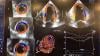
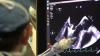
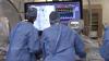
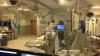
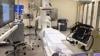
This is the newest cardiac cath lab at the University of Colorado Hospital in Aurora, Colorado. Construction was completed in June 2018. It is centered around a Philips Azurion Clarity IQ angiography system, which was chosen because its low X-ray dose imaging and guidance technologies that enable more complex, longer procedures. The room is use for the most involved complex percutaneous coronary interventions (PCI), including chronic total occlusions (CTOs) and complex high-risk indicated procedures (CHIP) patients. It is also used for transcatheter aortic valve replacement (TAVR), septal occluder procedures, transcatheter LAA closures and alcohol ablations.

The room is equipped for radial access procedures, which is used in a little more than 50 percent of cases at the hospital. It is also equipped with an Impella hemodynamic support system, wires and microcatheters for CTOs, and a SonoSite point of care ultrasound console for vascular access needle guidance.

See a 360 Degree View During a CTO Case in this room

Watch the VIDEO: The Evolution of Complex PCI at University of Colorado, which speaks with two of the operators who use this room.


Find more content from the University of Colorado Hospital.


EP Lab | January 03, 2019

This is a quick video tour of one of the dedicated electrophysiology (EP) labs at the University of Colorado Hospital. The room is built around a Siemens Artis Q.zen bi-plane angiography system. The system allows low radiation fluoro imaging, which reduced exposure to both patients and operators during long EP ablation procedures. 

Equipment in stock in this lab includes the Medtronic Artic Front cryoablation balloon.

Watch the VIDEO: Cryoballoon Ablation Best Practice Guidelines, an interview with Wilber Su, M.D.

Find more articles and videos on the University of Colorado Hospital

December 12, 2018

This is a walk around of the new Spectrum Dynamics Veriton SPECT-CT nuclear imaging system introduced at the 2018 Radiological Society of North America (RSNA) meeting.

This is a walk around of an innovative new SPECT-CT nuclear imaging system shown at the Radiological Society Of North America (RSNA) 2018 meeting this week. It's CT system with comes in 16, 64 or 128 slice configurations. It has 12 SPECT detector robotic arms that automatically move toward the patient and use a sensor to stop a few millimeters from the skin to optimize photon counts and SPECT image quality. It also uses more sensitive CZT digital detectors, which allows either faster scan times, or use of only half the radiotracer dose of analog detector scans.

Read the article “Nuclear Imaging Moves Toward Digital Detector Technology.” 

Read the article "Spectrum Dynamics Sues GE for Theft, Misappropriation of Trade Secrets and Unfair Competition."


Cardiovascular Ultrasound | December 12, 2018
This is an example of the new Fetal HQ heart and vascular software from GE Healthcare for fetal ultrasound. The software, for the Voluson E10, helps evaluate the fetal heart shape, size and contractibility. A feature called Radiant Flow shows the blood flow in a 3-D view. It can also help show slow-flow blood, such as neuro-vascular circulation. This was shown for the first time at the 2018 Radiological Society of North America (RSNA) meeting.


Cybersecurity | December 12, 2018

Anton S. Becker, M.D., radiology resident at the University Hospital of Zurich, Switzerland, discusses the long-term risks of cyberattacks on medical imaging data at the 2018 Radiological Society of North America (RSNA) annual meeting.

Advanced Visualization | December 12, 2018

This is an example of the FDA-cleared OpenSight augmented reality (AR) system for surgical planning from NovaRad at the Radiological Society Of North America (RSNA) 2018 meeting. It uses a HoloLens headset to register an overlay of the patient’s MR or CT scan in the patient. The operator can use hand movements to slice through and manipulate the images.

The vendor gained an FDA indication for AR to be used in surgical planning in 2018. NovaRad is working with the FDA for a second indication for use of the AR in the operating room during procedures.

This video is jerky, slightly misaligned and the hand movements did not always respond because itwas shot with an iPhone inside the visor. The image quality and hand movements are much better when actually wearing it on your head and aligned for the specific user.

Read more about this technology

Computed Tomography (CT) | November 14, 2018

Matthew Budoff, M.D., professor of medicine, David Geffen School of Medicine, UCLA, spoke at the 2018 American Heart Association (AHA) meeting on the new AHA guidelines for cholesterol, including the use of computed tomography (CT) calcium scoring scans to assess patient risk. Budoff has been involved in several of the coronary artery calcium (CAC) trials that supported the new recommendation.


Related CT Calcium Scorining Content:

VIDEO: New Cholesterol Guidelines Support CT Calcium Scoring for Risk Assessment — Interview with Matthew Budoff, M.D.

CT Calcium Scoring Becoming a Key Risk Factor Assessment

ACC and AHA Release Updated Cholesterol Guidelines for 2018

VIDEO: CT Calcium Scoring to Screen For Who Should Take Statins — Interview with Matthew Budoff, M.D.

VIDEO: The History of CT Calcium Scoring — Interview with Arthur Agatston, M.D.


Subscribe Now