Videos

Cardiac Imaging

Congenital Heart | November 01, 2018

Example of GE Healthcare’s FetalHQ software for the ultrasound imaging of fetal hearts. The new tool runs on GE Healthcare’s Voluson ultrasound systems and is the first tool to simultaneously examine the size, shape and function of the fetal heart echocardiography for congenital heart evaluations.

Read more about the technology in the article "Cardiac Ultrasound Software Streamlines Fetal Heart Exams."


TCT | October 03, 2018

DAIC Editor Dave Fornell takes a tour of some of the most innovative new cardiovascular technology he found on the expo floor at the 2018 Transcatheter Cardiovascular Therapeutics (TCT) conference.

Here are links to other Editor's Choice videos at recent conferences:

VIDEO: Editor's Choice of Most Innovative New Cardiac Technology at ACC 2018

VIDEO: Editor’s Choice of the Most Innovative Echo Technology at ASE 2018

VIDEO: Editor's Choice of the Most Innovative New Imaging Technology at RSNA 2017


#TCT2018

Artificial Intelligence | September 27, 2018

Rami Doukky, M.D., professor of medicine, preventative medicine and radiology, and chief of the Division of Cardiology at Cook County Health and Hospitals System, discusses how artificial intelligence (AI) will impact all medical imaging modalities at the 2018 American Society of Nuclear Cardiology (ASNC) annual meeting in San Francisco.


Related Artificial Intelligence Content in Cardiac Imaging:

VIDEO: Artificial Intelligence in Cardiac Ultrasound — Interview with Partho Sengupta, M.D.

How Artificial Intelligence Will Change Medical Imaging

Combatting the World’s No. 1 Cause of Death With the Help of Artificial Intelligence and Advanced Technology

Advances in Cardiac Imaging Technologies at RSNA 2017


Cardiac Imaging | September 10, 2018

Leslee Shaw, Ph.D., professor of cardiology and radiology, Weill Cornell Medical Center, New York, and past-president of the Society of Cardiovascular Computed Tomography (SCCT), explains how cardiac imaging will be used in the ISCHEMIA Trial to assess treatment strategies for patients with stable ischemic heart disease. The trial was discussed by Shaw at the 2018 American Society of Nuclear Cardiology (ASNC) meeting.

Watch the related VIDEO Role of Cardiac CT in Value-based Medicine, an interview with Shaw at SCCT 2017.

Nuclear Imaging | August 22, 2018

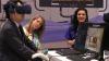
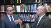
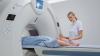
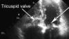
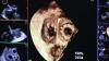
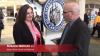
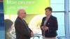
Kim A. Williams, Sr., M.D., MACC, MASNC, FAHA, FESC, cardiology division chief and James B. Herrick professor at Rush University Medical Center, discusses the importance of nuclear cardiology in preventive medicine, and previews his upcoming keynote lecture at the 2018 annual meeting of the American Society of Nuclear Cardiology (ASNC), Sept. 6-9 in San Francisco.

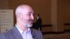
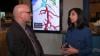
Watch the related VIDEO ASNC 2018 Program Preview, where Rami Doukky, M.D., professor of medicine, preventive medicine and radiology, and chief of the Division of Cardiology at Cook County Health and Hospitals System, discusses new additions to the ASNC meeting program for 2018.

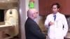
Watch the VIDEO MACRA's Impact on Cardiology, an interview with Williams on the impact of healthcare reform on cardiology and specifically on nuclear perfusion imaging.

Nuclear Imaging | August 21, 2018

Rami Doukky, M.D., professor of medicine, preventive medicine and radiology, and chief of the Division of Cardiology at Cook County Health and Hospitals System, discusses new additions to the program for the 2018 annual meeting of the American Society of Nuclear Cardiology (ASNC), Sept. 6-9 in San Francisco.

Watch the VIDEO: Imaging in the Assessment of Preventive Cardiology — interview with Kim Williams, M.D., about his key note lecture at the 2018 ASNC.

Watch the VIDEO: American Society of Nuclear Cardiology (ASNC) Celebrates 25 Years — interview with Robert C. Hendel, M.D.

Watch the VIDEO Clinical Decision Support Requirements for Cardiac Imaging, an interview with Doukky at the 2017 ASNC Today meeting.

Cath Lab | August 13, 2018

Jeffrey Schussler, M.D., FACC, FSCAI, FSCCT, FACP, interventional cardiologist at Baylor Scott White Heart and Vascular Hospital, Dallas, explains some of the key innovations his institution is using to improve cardiovascular care. These include the use of robotic percutaneous coronary intervention (PCI), FFR-CT imaging and transcatheter valve technologies.

Take a 360 photo look inside a Baylor combo cardiac cath/EP lab. 

Watch the VIDEO: Dedicated Cardiac MRI Use at the Baylor Scott White Heart Hospital — an interview with Haojie Wang, M.D., director of advanced cardiovascular MRI and a member of the heart valve clinic at Baylor Scott White Heart Hospital, Dallas.


Magnetic Resonance Imaging (MRI) | August 13, 2018

Haojie Wang, M.D., director of advanced cardiovascular MRI and a member of the heart valve clinic at Baylor Scott White Heart Hospital, Dallas, explains the utility of magnetic resonance imaging (MRI) in cardiology and the implementation of the cardiac MRI program at his hospital.

See a 360 degree photo of the Baylor MRI control room.

Watch the VIDEO: Implementation of New Cardiovascular Technologies at Baylor Scott White Heart Hospital — interview with Jeffrey Schussler, M.D., FACC, FSCAI, FSCCT, FACP, interventional cardiologist at Baylor Scott White Heart and Vascular Hospital, Dallas.
 

Watch the VIDEO: Advances in Cardiac MRI Technology — Interview with James Carr, M.D.

ASNC | July 25, 2018

Robert C. Hendel, M.D., FACC, FAHA, MASNC, director, Tulane Heart and Vascular Institute, explains the impact the ASNC has had on cardiac nuclear imaging during this anniversary year. 

The Annual Scientific Session of the American Society of Nuclear Cardiology is Sept. 6-9, 2018, at the San Francisco Marriott Marquis.

#ASNC2018

Computed Tomography (CT) | July 25, 2018

A discussion with Patricia Dickson, LRT(CT), director of imaging and outpatient services, Capital Cardiology Associates, Albany, N.Y., and Nikki Weber, RT(R)(CT)(M), a lead CT technologist at the Mayo Clinic, Rochester, Minn. They presented in sessions at the Society of Cardiovascular Computed Tomography (SCCT) 2018 meeting.


Related Tips to Improve CT Imaging Content:

VIDEO: Tips and Tricks to Aid Cardiac CT Technologist Workflow

VIDEO: How to Prepare a Patient for a Cardiac CT Scan

VIDEO: 10 Tips to Improve Cardiac CT Imaging

Find more SCCT news and videos


Computed Tomography (CT) | July 23, 2018

Ed Nicol, M.D., FSCCT, MBA, head of cardiac CT, Royal Brompton Hospital, London, and chair of the Society of Cardiovascular Computed Tomography (SCCT) Education Committee. He outlined new initiatives by SCCT to expand its educational offerings. For more information on SCCT's online educational links go to https://scct.org/page/Education.

Link to the technologist video referenced in this interview that is among the most viewed videos on DAIC — VIDEO: How to Prepare a Patient for a Cardiac CT Scan.

CT Angiography (CTA) | July 19, 2018

Kavitha Chinnaiyan, M.D., FACC, FSCCT, associate professor, Oakland University, William Beaumont School of Medicine, Royal Oak, Mich. She presented at the Society of Cardiovascular Computed Tomography (SCCT) 2018 meeting. 

She explains how noninvasive fractional flow reserve computed tomography (FFR-CT) is being used at Beaumont to assess patients presenting with chest pain. FFR-CT has been used to triage these patients faster than doing a series of troponin tests, keeping patients overnight for a nuclear scan or sending them for a diagnostic angiogram. Most patients can be discharge is a fraction of the time compared to these other standards of care. Chinnaiyan also showed examples of FFR-CT, CT angiograms and cath lab invasive angiograms of the same lesions to show the correlation between the different modalities. Some examples show what appears to be a severe lesion, but FFR-CT shows acceptable blood flow past lesions so no revascularization was required.


Related FFR-CT Content:

Clinical Applications of FFR-CT

VIDEO: Implementation and the Science Behind FFR-CT — interview with James Min, M.D.

VIDEO: Early U.S. Experience With FFR-CT in Evaluating ED Chest Pain Presentation — interview with Simon Dixon, M.D.

VIDEO: Status of FFR-CT Adoption in the United States — interview with Campbell Rogers, M.D.


July 18, 2018

Suhny Abbara, M.D., FSCCT, chief of cardiothoracic imaging and chair of the CT operations committee, University of Texas Southwestern Medical Center, Dallas, incoming president of the Society of Cardiovascular Computed Tomography (SCCT), and editor of the new RSNA journal Radiology: Cardiothoracic Imaging. He spoke to DAIC at SCCT 2018 meeting. 

Read the related article "Spectral Imaging Brings New Light to CT."


CT Angiography (CTA) | July 18, 2018

A discussion with Gianluca Pontone, M.D., Ph.D., FSCCT, director of cardiovascular MRI, Centro Cardiologico Manzino, Milan, Italy, at the Society of Cardiovascular Computed Tomography (SCCT) 2018 meeting. He said CT can be comparable to the gold-standards of nuclear and MRI perfusion depending on the scanner used to acquire the images. 

Read the article "CT Perfusion Imaging Ready for Mainstream." 

Heart Valve Technology | July 18, 2018

Jonathon Leipsic, M.D., FSCCT, professor of radiology and cardiology at the University of British Columbia, Vancouver, Canada, and an expert in transcatheter valve imaging. He spoke about his experiences with TAVR and TMVR trials and devices planning at the the Society of Cardiovascular Computed Tomography (SCCT) 2018 meeting.

Read the related article "The Essentials of Structural Heart Imaging."

Watch the VIDEO "What to Look for in CT Structural Heart Planning Software."


Computed Tomography (CT) | July 17, 2018

An interview with Patrick Serruys, M.D., Ph.D., Imperial College London, principal investigator of the SYNTAX III Trial presented earlier this year as a late-breaker at EuroPCR. He presented the trial again at the Society of Cardiovascular Computed Tomography (SCCT) 2018 meeting. 

He explains how noninvasive fractional flow reserve computed tomography (FFR-CT) was used in the study and compared to invasive angiography and pressure wire based FFR. Serruys, a well-known pioneer in interventional cardiology, had high praise for FFR-CT after seeing it in real-world cases. He said the technology can be used to better select patients who undergo cardiac CT scans to determine who needs to be revascularized in the cath lab. This can reduce the current issue where a lot of patients who have antomical diease on CT imaging turn out to not have a severe enough lesion to require stenting.

Read the article "SYNTAX III Revolution Trial Shows CT-FFR Could Replace Cine-angiography in Coming Years."

CT Angiography (CTA) | July 17, 2018

A discussion with Todd Villines, M.D., FACC, FAHA, FSCCT, director of cardiovascular research and cardiac CT programs at the Walter Reed National Military Medical Center in Bethesda, Md., and president of the Society of Cardiovascular Computed Tomography (SCCT). He outlines key technology trends at the 2018 SCCT meeting.


Related CT Technology Advances Content:

New Cardiovascular CT Technology Entering the Market

PODCAST: Shortcomings of CTA in Cardiology — Interview with Todd C. Villines, M.D.,

VIDEO: Editor's Choice of Most Innovative New Cardiac CT Technology

VIDEO: Tips and Tricks to Aid Cardiac CT Technologist Workflow

VIDEO: Cardiovascular CT Educational Initiatives by SCCT — Interview with Ed Nicol, M.D.

VIDEO: Using FFR-CT in Everyday Practice — Kavitha Chinnaiyan, M.D.

VIDEO: Applications of Spectral CT — Interview with Suhny Abbara, M.D.

VIDEO: The Essentials of CT Transcatheter Valve Imaging — Interview with Jonathon Leipsic, M.D.

VIDEO: The State of CT Myocardial Perfusion Imaging — Interview with Gianluca Pontone, M.D.


Cardiovascular Ultrasound | July 10, 2018

DAIC Editor Dave Fornell highlights some of the most innovative new technology on the show floor of the American Society of Echocardiography (ASE) 2018 meeting. The segments include virtual reality workstations, advanced 3-D cardiac ultrasound quantification and visualization, improved echo-fluoro image fusion technology, and imaging aided by artificial intelligence.


Artificial Intelligence | July 10, 2018

Partho Sengupta, M.D., DM, FACC, FASE, chief division of cardiology, director of cardiac imaging, West Virginia University Heart and Vascular Institute, explains how artificial intelligence is being integrated into echocardiography and used to mine big data to better assess patients at the American Society of Echocardiography (ASE) 2018 meeting. 

Watch the VIDEO: Ultrasound's Integration of Artificial Intelligence and Robotic Echo, with Senguta at the ASE 2017 meeting


Heart Valve Technology | July 05, 2018

Rebecca Hahn, M.D., professor of medicine and director of interventional echocardiography, Columbia University Medical Center, New York, explains that techniques for imaging the tricuspid valve, the  advanced study of its disease etiology and the rapid development of transcatheter devices to treat tricuspid valve disease are all developing together. She spoke on these topics during sessions at both the American Society of Echocardiography (ASE) 2018 meeting and at the Transcatheter Valve Therapies (TVT) conference in June. 


Related Transcatheter Tricuspid Valve Content:

Recent Advances in Transcatheter Valve Technology

VIDEO: Mitral and Tricuspid Valve Repair Technologies — interview with Azeem Latib, M.D.

VIDEO: Transcatheter Tricuspid Valve Repair and Replacement Technologies — interview with Rebecca Hahn, M.D.

Minneapolis Heart Institute Foundation Enrolls First Patient in TRILUMINATE Tricuspid Repair Trial

Positive, Sustained Improvement at One Year in SCOUT I Transcatheter Tricuspid Repair Trial

Gate Bioprosthesis Used in Canada's First Transcatheter Valve Replacement for Tricuspid Regurgitation

Cardiovascular Ultrasound | July 03, 2018

Michael Lanspa, M.D., director of critical care echocardiography services, Intermountain Medical Center, Salt Lake City, Utah, explains the training initiatives by the American Society of Echocardiography (ASE) for point-of-care ultrasound. Lanspa serves on the educational committees for ASE, the American Thoracic Society (ATS) and the Society of Critical Care Medicine. 

POCUS has seen a big expansion in cardiology to better assess patients before sending them for more advanbced imaging or tests. POCUS is also seeing widespread expansion in many other areas of healthcare, especially in emergency medicine.

POC ultrasound was discussed as one of the big recent developments at ASE 2018

VIDEO: Imaging COVID-19 With Point-of-Care Ultrasound (POCUS)

VIDEO: Automated Cardiac Ejection Fraction for Point-of-care-ultrasound Using Artificial Intelligence


Cardiovascular Ultrasound | July 02, 2018

Lissa Sugeng, M.D., associate professor of medicine, director of echocardiography and of the Yale Echo Core Lab, Yale School of Medicine, explains the advances and advantages of using 3-D cardiac ultrasound during the American Society of Echocardiography (ASE) 2018 meeting. She said 3-D echo still has slower frame rates than 2-D, but offers some advantages. Sugeng said echo labs should consider adding at least 3-D machine, as this technology will become the way of the future in echo. Read more about recent advances in cardiac ultrasound. 

Cardiovascular Ultrasound | July 02, 2018

This is an example of the a mitral valve with regurgitation imaged with 3-D echocardiography using the Siemens SC2000 cardiac ultrasound system. The first video clip shows the 3-D anatomy of the heart, the mitral valve, its leaflet cusps tabled and the left ventricular outflow track (LVOT) of the aortic valve. The second clip shows how the software can automatically extract and quantify the regurgitant volume. The last clip shows an isolation of the mitral valve and the regurgitant jet, showing the exact location of the source, which can be used aid surgical planning. This video was shot at the American Society of Echocardiography (ASE) 2018.

Find additional technology video clips, video interviews and news from ASE.


Cardiovascular Ultrasound | June 28, 2018

Sunil Mankad, M.D., FASE, ASE 2018 meeting program chair and director of transesophageal echocardiography at Mayo Clinic, Rochester, Minn., explains four big trends in cardiac ultrasound during the American Society of Echocardiography (ASE) 2018 meeting. These include the advancement of 3D echo, point-of-care ultrasound, artificial intelligence and new forms of image visualization. Read more about recent advances in cardiac ultrasound. 

Cardiovascular Ultrasound | June 28, 2018

This is a demonstration of the the Philips TrueVue photo-realistic rendering and lighting source technology. This example highlights a transcatheter ASD closure with two Amplatzer occluders. The technology allows users to changing the lighting conditions to improve contrast. The light source can also be moved around to change the shadows and add more depth perception. The light source also can be pushed through the tissue to back light the anatomical structures, as shown in this example at the American Society of Echocardiography (ASE) 2018 meeting. The backlighting confirms a gap at the top of the image where part of the septal defect remains exposed. 

Find more videos and news from ASE 

Cardiovascular Ultrasound | June 11, 2018

Thomas Porter, M.D., professor of medicine and director of the echocardiography lab at the University of Nebraska Medical Center, discusses potential therapeutic uses for microbubble contrast ultrasound at the 2017 American Society of Echocardiography (ASE) annual meeting.

Cath Lab | March 27, 2018

Roxana Mehran, M.D., FACC, FACP, FCCP, FESC, FAHA, FSCAI, professor of medicine and director of interventional cardiovascular research and clinical trials at the Zena and Michael A. Wiener Cardiovascular Institute at Mount Sinai School of Medicine. She spoke on how to avoid acute kidney injury (AKI) and contrast-induced nephropathy (CIN) using various strategies in the cath lab to reduce iodine contrast use at the American College of Cardiology (ACC) 2018 meeting. 


Related CIN Content:

Sliding Scale Hydration Washes Out Contrast-Induced Kidney Complications

Contrast-Induced Kidney Injury Prevented With RenalGuard in REMEDIAL III Trial

VIDEO: How to Avoid Acute Kidney Injury in the Cath Lab — Interview with Hitinder Gurm, M.D.

VIDEO: Combating Contrast Induced Nephropathy in the POSEIDON Trial — Presentation by Sonjot Brar, M.D.


ACC | March 21, 2018

DAIC Editor Dave Fornell takes a tour of some of the most interesting new technologies on the expo floor at the 2018 meeting of the American College of Cardiology (ACC). Find links to other videos and the ACC 2018 Late-Breaking Trials.


Cath Lab | March 15, 2018

A discussion with Hitinder Gurm, M.D., MBBS, FACC, professor, internal medicine, and associate chief clinical officer, University of Michigan Health System, at the 2018 American College of Cardiology (ACC) meeting.  For more information, read the article "Understanding Contrast-Induced Nephropathy." He discusses various strategies to help reduce iodine contrast use in the cath lab to better protect patients from CIN.


Related CIN Content:

Sliding Scale Hydration Washes Out Contrast-Induced Kidney Complications

Contrast-Induced Kidney Injury Prevented With RenalGuard in REMEDIAL III Trial

VIDEO: Strategies to Avoid Acute Kidney Injury Caused by Cath Lab Contrast — Interview with Roxana Mehran, M.D.

VIDEO: Combating Contrast Induced Nephropathy in the POSEIDON Trial — Presntation by Sonjot Brar, M.D.


Mobile Devices | February 16, 2018

DAIC Editor Dave Fornell previews the launch of augmented reality (AR) technology in the March/April 2018 issue of DAIC. Augmented reality brings new depth to print content through your smartphone by connecting to related videos and other resources.

Cardio-oncology | February 01, 2018

American Heart Association (AHA) released a scientific statement Feb. 1, 2018, linking breast cancer and cardiovascular disease. Laxmi Mehta, M.D., author of the scientific statement and director of the Women’s Cardiovascular Health Program at The Ohio State University Wexner Medical Center, discusses a case from her center as an example of how the subspecialty of cardio-oncology is helping patients.

Read the article “AHA Puts New Focus on Where Heart Disease and Breast Cancer Treatment Meet.”

Watch the "VIDEO: Assessing Cardiotoxicity Response With Cardio-Oncology Echo Imaging."
 

Cardiac Imaging | December 29, 2017

ITN and DAIC Editor Dave Fornell takes a tour of some of the most interesting new medical imaging technologies on the expo floor at the 2017 Radiological Society of North America (RSNA) meeting. Watch the editor’s choice video for new health IT technologies at HIMSS 2017, new advances in cardiac ultrasound at ASE 2017, and CT advances at the SCCT 2017 meeting.

Magnetic Resonance Imaging (MRI) | December 13, 2017

Emanuel Kanal, M.D., director of MRI services and professor of radiology and neuroradiology at the University of Pittsburgh Medical Center, explains the new mobile application he developed, which lets users visually model the forces at work during an MRI exam on patients with implanted medical devices, at the 2017 Radiological Society of North America (RSNA) meeting.

Read the related article “The Changing Relationship Between MRIs and Pacemakers.”

Advanced Visualization | December 07, 2017

Dianna Bardo M.D., director of body MR and co-director of the 3-D Innovation Lab at Phoenix Children's Hospital, discusses how 3-D printing and other advanced imaging modalities can help improve outcomes in complex cases. Read the article “The Use of 3-D Printing in Cardiology.” Watch the WEBINAR “Innovation and Success in 3D-inspired Development of the Business and Clinical Practice,” presented by Bardo.


Angiography | November 22, 2017

Shimadzu demonstrated its Trinias Interventional X-ray System at the 2017 Transcatheter Cardiovascular Therapeutics (TCT) conference. The system offers several features to enable improved patient access and procedural guidance at a lower radiation dose than previous generation angiography systems. For more information visit www.shimadzu.com/med/products/angio.

Medical 3-D Printing | November 17, 2017

Dee Dee Wang, M.D., director, structural heart imaging at Henry Ford Hospital, Detroit, explains how her center uses 3-D printing and computer aided design (CAD) software to improve patient outcomes. She spoke to DAIC at the 2017 Transcatheter Cardiovascular Therapeutics (TCT) annual meeting. Read the articles “The Use of 3-D Printing in Cardiology” and “Henry Ford Hospital Study Shows 3-D Imaging Improves Fixing Broken Hearts.”  

Additional articles and videos on Henry Ford Hospital 

TCT | November 15, 2017

DAIC Editor Dave Fornell shows some of the innovations displayed on the expo floor at the 2017 Transcatheter Cardiovascular Therapeutics (TCT). Hear what Cardiovascular Research Foundation (CRF) President and CEO Juan Granada, M.D., feels are the hottest technologies to watch. 

Cardiovascular Ultrasound | October 12, 2017

Sharon Mulvagh, M.D., FRCPC, FACC, FAHA, FASE, professor, Division of Cardiology, Department of Medicine, Dalhousie University, Nova Scotia Health Authority, Halifax, Canada, and emeritus professor at Mayo Clinic, discusses how ultrasound can be used to assess plaque burden at the 2017 American  Society of Echocardiography (ASE) annual meeting. Read the article highlighting new technologies at ASE 2017 "A Glimpse Into the Future of Cardiac Ultrasound." Watch her presentation on the use of point-of-care echo for plaque burden risk assessment from ASE 2017.


Cardiovascular Ultrasound | August 30, 2017

This video educational session, provided in partnership with the American Society of Echocardiography (ASE), is titled "Vascular Ultrasound in Subclinical Disease." It is presented by Valentin Fuster, M.D., Ph.D., chief of the Division of Cardiology and director of Mount Sinai Heart, the Zena and Michael A. Wiener Cardiovascular Institute, and the Marie-Josée and Henry R. Kravis Center for Cardiovascular Health at Icahn School of Medicine at Mount Sinai.. This is one of the sessions presented at the ASE 2016 annual scientific sessions, one of the premier meetings for cardiac ultrasound education. For more information, visit asescientificsessions.org. 

For more educational videos on echocardiography, visit the DAIC education channel. 

Subscribe Now