Videos

VIDEO: One on One with Hal Wolf, FHIMSS, HIMSS President and CEO

HIMSS | March 05, 2024

Industry trade shows and conferences seem to be making their comeback in 2024. And the Healthcare Information and Management Systems Society (HIMSS) Global Conference and Exhibition seems particularly poised to deliver the best of the best when it comes to digital transformation in both the delivery of healthcare, but also the delivery of a quality experience for those in this demanding, rapidly evolving industry. This month in our ongoing One on One series with industry leaders, we are talking with Hal Wolf, FHIMSS, president and CEO of HIMSS. He offered insights on the society’s new partnership with Informa Markets, key topics being covered at HIMSS24, AI’s impact on the industry, and his thoughts on healthcare sustainability.

Find more HIMSS24 conference coverage here

A New Partnership for Growth

Last August, Informa Markets and HIMSS announced a landmark partnership to propel the growth and evolution of the HIMSS Global Health Conference and Exhibition, recognized as the most influential healthcare technology event of the year, and in North America. It draws 40,000 health professionals, tech leaders, providers and governmental organizations from across the globe. Informa Markets, the world’s largest exhibition organizer, took on management of the HIMSS Exhibition, while HIMSS continues to oversee developing expert content and programming.

Exciting New Features at HIMSS24

At HIMSS2024, with this new collaboration comes new features, including:

Related content:

Find more HIMSS24 conference coverage here

HIMSS Launches Modernized Infrastructure Adoption Model to Support Global Digital Health Transformation

Top Public Policy Experts at HIMSS24 to Address Global AI Landscape and Digital Transformation in Healthcare

VIDEO: Using Maturity Models to Measure Digital Health

VIDEO: Moving Digital Transformation Forward in Healthcare

VIDEO: Key Components to Creating and Implementing AI and Digital Transformation Solutions

VIDEO: The Benefits and Pitfalls of Artificial Intelligence in Healthcare

VIDEO: A Look at Cybersecurity and How Healthcare is at Risk

Information Technology

September 27, 2019

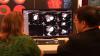
Akshay Khandelwal, M.D., director of medical operations at the Henry Ford Heart and Vascular Institute, Detroit, and associate professor of cardiology at Wayne State University, explains how his center has reduced X-ray radiation dose in the cardiac catheterization labs.

Watch the related VIDEO: Reducing Cath Lab Radiation Dose at Henry Ford Hospital — Discussion with Nicolas Bevins, Ph.D., vice chair, physics and research, and Jessica Harrington, RCIS, Henry Ford Hospital.

Additional articles and videos on Henry Ford Hospital 


For more on how to reduce dose in the cath lan, read these related articles:

Cardiology Societies Call for Better Radiation Dose Tracking

Defining the Cath Lab Workplace Radiation Safety Hazard

Dose-Lowering Practices for Cath Lab Angiography

5 Technologies to Reduce Cath Lab Radiation Exposure

VIDEO: Heart Surgeon Shares Effects of Fluoroscopic Radiation Exposure

Helping Interventional Cardiologists Reduce Exposure to Ionizing Radiation

14 Ways to Reduce Radiation Exposure in the Cath Lab


(Editor's note - this video was originally published in November 2018 and was revised September 2019)


Advanced Visualization | August 09, 2019

An example of Siemens' photo-realistic Cinematic image reconstruction. This image is from a CTA exam from a Siemens Force CT scanner. 

Vendors who offer this realistic type of CT image rendering say it is not used for diagnostics. However, the technology can be helpful when explaining things to the patient and their family, educating physicians and staff, and for surgeons, since it offers a realistic view of the anatomy that is easier for most people to understand who are not familiar with cardiac anatomy as it appears in traditional CT multiplanar reconstruction (MPR) images. 

This example of software was demonstrated on the expo floor at the 2019 Society of Cardiovascular Computed Tomography (SCCT) meeting. 


Find more news and videos from the SCCT meeting

CT Angiography (CTA) | August 08, 2019

This is a quick video example of a cardiac computed tomography (CT) exam showing a Medtronic CoreValve transcatheter aortic valve replacement (TAVR) device implanted. The image was reconstructed using Canon Medical’s Global Illumination photo-realistic rendering advanced visualization post-processing software. Vendors who offer this realistic type of CT image rendering say it is not used for diagnostics. However, the technology can be helpful when explaining things to the patient and their family, educating physicians and staff, and for surgeons, since it offers a realistic view of the anatomy that is easier for most people to understand who are not familiar with cardiac anatomy as it appears in traditional CT multiplanar reconstruction (MPR) images. 

This example of software was demonstrated on the expo floor at the 2019 Society of Cardiovascular Computed Tomography (SCCT) meeting. 


Heart Valve Technology | July 24, 2019

Joao Cavalcante, M.D., FSCCT, director of structural heart CT and cardiac MRI, Minneapolis Heart Institute, discusses transcatheter mitral valve replacement (TMVR) imaging requirements at the Society of Cardiovascular Computed Tomography (SCCT) 2019 meeting. SCCT held its first TMVR planning hands-on workshop, which he was involved in. TMVR is expanding with currently FDA cleared valve-in-valve (VIV) and valve-in-ring (VIR) procedures.


Related TMVR Content:

Interventional Imagers: The Conductors of the Heart Team Orchestra

VIDEO: The Importance of the Neo-LVOT in Transcatheter Mitral Valve Replacement  — Interview with Dee Dee Wang, M.D.

VIDEO: Transcatheter Structural Heart Procedure Navigation Technology Advances  — Interview with Stephen Little, M.D.

Recent Advances in Transcatheter Valve Technology

Abbott Begins Tendyne Transcatheter Mitral Valve U.S. Pivotal Trial

VIDEO: The Essentials of CT Transcatheter Valve Imaging — Interview with Jonathon Leipsic, M.D.,

New LAMPOON Technique Reduces LVOT Obstruction in Transcatheter Mitral Valve Replacement

Find more SCCT news and videos

Cardiovascular Ultrasound | July 10, 2019

This is an example of a cardiac echocardiography exam performed using an iPhone and the Butterfly IQ ultrasound transducer and app. The company is exhibiting at the American Society Of Echocardiography (ASE) 2019 meeting. This is the first “ultrasound system on a chip.” The Butterfly IQ ultrasound system consists of a transducer that connects to an iPhone or iPad to record ultrasound exams. The system has 18 different applications for specialized images, including cardiac imaging, vascular, lung, abdominal and others. The apps allow for quantification and offers many of the features of larger cart-based systems. The company is working on incorporating artificial intelligence technologies to automate some processes, such as auto ejection fractions.

This technology was picked as one of the big ultrasound advances in the VIDEO: 4 Recent Advances in Echocardiography Technology and in the VIDEO: Editor's Choice of the Most Innovative New Technology at RSNA 2018.

Artificial Intelligence | July 02, 2019

Judy Hung, M.D., director of echocardiography, Division of Cardiology, Massachusetts General Hospital, Boston, discusses how her center is partnering to develop artificial intelligence (AI) applications to help improve image quality and speed workflow in cardiac ultrasound. She spoke to DAIC at the 2019 American Society of Echocardiography (ASE).


Cardiovascular Ultrasound | July 01, 2019

Partho Sengupta, M.D., MBBS, chief of cardiology, West Virginia Heart and Vascular Institute, explains how wearable, smartphone-based apps and medical devices, and artificial intelligence (AI) might be used to cost-effectively triage larger numbers of patients in rural areas and in the developing world for serious diseases. He spoke at the 2019 American Society Of Echocardiography (ASE) meeting.

Sengupta is involved with a pilot program using the Butterfly app and transducer to turn a smartphone into an inexpensive ultrasound system. He said the idea is to have novice ultrasound users screen more patients with these types of devices and the exams either being sent to a remote hospital for reading. He said AI algorithms also could be used to help flag any exams that show abnormalities, which would greatly speed reads and getting these patients treatment. 

Watch the related VIDEO: How Smartphones May Revolutionize Healthcare in the Developing World — Interview with Jacques Kpodonu, M.D.,

Find more news and video from ASE 2019


Artificial Intelligence | June 28, 2019

This is a quick example of how artificial intelligence (AI) is being integrated on the back end of cardiac ultrasound systems to help automate and speed workflows. This video segment is from the expo floor of the 2019  American Society of Echocardiography (ASE) meeting. It shows AI-based AFI automation features incorporated into the GE Vivid E95 echo system. 

Read a blog about how AI will help advance cardiac ultrasound imaging, Combatting the No. 1 Cause of Death With the Help of Artificial Intelligence and Advanced Technology.

Find more ASE 2019 coverage


Cardiovascular Ultrasound | June 28, 2019
Virtual and Augmented Reality | June 27, 2019

Roberto Lang, M.D., director of cardiac imaging at the University of Chicago, has been working with TomTec for the past three years on a project to use virtual reality (VR) to edit and view 3-D cardiac ultrasound and CT scans. He spoke at a couple sessions this week at the 2019 American Society Of Echocardiography (ASE) meeting, where doctors were able to view the session using VR headsets. The hands-on demonstration of this technology in the TomTec booth at ASE 2018 and 2019 was one of the most popular exhibits with attendees both years.

Read more about this technology in Top Technology Trends in Echocardiography at ASE 2018

See examples of VR technology for echo in the  VIDEO: Editor’s Choice of the Most Innovative Echo Technology at ASE 2018

Here is a video example of this technology being demonstrated at ASE19.


Wearables | June 21, 2019

Jacques Kpodonu, M.D., FACC, cardiac surgeon, Beth Israel Deaconess Medical Center, and professor at Harvard Medical School, explains how medical devices and wearables that interface with smartphones and apps might be used to eliminate healthcare disparities in rural areas of the developed world and help raise the level of care in the developing world. He spoke at the 2019 AI-Med Cardiology conference. 


Related Smartphone and Wearable Content:

VIDEO: Use of Wearable Medical Devices for Cardiac Rehabilitation — Interview with Robert Klempfner, M.D.

VIDEO: Use of Wearables to Track Electrophysiology Patients — Interview with Khaldoun Tarakji, M.D.

Smartphones Used to Successfully Screen More than 60,000 for Atrial Fibrillation

VIDEO: The Future of Wearables in Healthcare — Interview with Karl Poterack, M.D.

VIDEO: Artificial Intelligence Applications for Cardiology — Interview with Anthony Chang, M.D.
 

Atrial Fibrillation | June 21, 2019

Sanjaya Gupta, M.D., electrophysiologist, St. Luke's Mid America Heart Institute, and assistant professor, University of Missouri–Kansas City School of Medicine, explains how his center developed an artificial intelligence (AI) application to automatically risk stratify atrial fibrillation (AFib) patients. The Epic-based app stratifies patients into those who should be placed on anticoagulation and those who are candidates for left atrial appendage (LAA) occlusion. He spoke at the 2019 AI-Med Cardiology conference. 

His center hopes to develop similar guidelines based AI apps for other types of cardiac risk scoring. Gupta said he is looking for other centers to partner with to co-develop and test these AI apps.    


Related Cardiology AI Content:

VIDEO: Overview of Artificial Intelligence and its Use in Cardiology — Interview with Anthony Chang, M.D.

VIDEO: ACC Efforts to Advance Evidence-based Implementation of AI in Cardiovascular Care — Interview with John Rumsfeld, M.D.

VIDEO: Artificial Intelligence Applications for Cardiology — Interview with Anthony Chang, M.D.

PODCAST: Fitting Artificial Intelligence Into Cardiology — Interview with Anthony Chang, M.D.

VIDEO: How Hospitals Should Prepare for Artificial Intelligence Implementation — Interview with Paul Chang, M.D.

Technology Report: Artificial Intelligence 

Artificial Intelligence | June 20, 2019

John Rumsfeld, M.D., Ph.D., FACC, American College Cardiology (ACC) chief innovation officer, and professor of medicine at the University of Colorado School of Medicine, explains how the ACC is working with artificial intelligence (AI) vendors to directly impact cardiac care. He said there is a tremendous amount of investment in and hype surrounding AI in healthcare, but to date there has been very little of this has translated in to changes in the way cardiology care is delivered. He outlines several areas to successfully apply AI to improve cardiovascular care and outcomes. He also discussed the current ACC efforts to advance evidence-based implementation of AI in cardiac care including applications for the NCDR.

He spoke at the 2019 Cardiology AI-Med conference. 

Watch the related VIDEO: Overview of Artificial Intelligence and its Use in Cardiology, an interview with Anthony Chang, M.D., chief artificial intelligence officer, Children’s Hospital of Orange County (CHOC), and founder of AIMed.  


Artificial Intelligence | June 18, 2019

Anthony Chang, M.D., chief artificial intelligence officer, Children’s Hospital of Orange County (CHOC), and founder, AIMed, explains the basic principles of artificial intelligence (AI) in medicine. He outlines some basic AI definitions, potential programming biases and use cases. He also explains the need for the Cardiology AI-Med conference, which held its inaugural meeting in June 2019 in Chicago.


Related AI Content:

Link to all the recorded AI-Med Cardiology conference sessions

VIDEO: Artificial Intelligence Applications for Cardiology — Interview with Anthony Chang, M.D.

PODCAST: Fitting Artificial Intelligence Into Cardiology — Interview with Anthony Chang, M.D.

VIDEO: How Hospitals Should Prepare for Artificial Intelligence Implementation — Interview with Paul Chang, M.D.

Technology Report: Artificial Intelligence 

Medical 3-D Printing | May 21, 2019

This is a sample of the 3-D printed hearts and coronary anatomy models created from patient CT scans to enable anatomical assessment, device sizing and plan which devices to use and navigation for complex structural heart cases at Henry Ford Hospital, Detroit, Mich. These hearts are in the office of Dee Dee Wang, M.D., director of structural heart imaging, at Henry Ford. She is in charge of a robust 3-D printing program to aid the structural heart program, which surpassed its 1,000th patient printed heart earlier in 2019. 


Realted Content With Wang:

VIDEO: Applications in Cardiology for 3-D Printing and Computer Aided Design

VIDEO: The Importance of the Neo-LVOT in Transcatheter Mitral Valve Replacement

Interventional Imagers: The Conductors of the Heart Team Orchestra


Additional articles and videos on Henry Ford Hospital 


Heart Valve Technology | May 20, 2019

A demonstration of how to calculate the neo-left ventricular outflow tract (neo-LVOT) on CT imaging for a transcatheter mitral valve replacement using Circle Imaging's advanced visualization software. The demonstration looks at the use of an Edward's Sapien valve being implanted for a mitral valve-in-valve procedure. The overhang of the Sapien can block the LVOT blood flow, which can be catastrophic for the patient. So, assessment of the neo-LVOT in a simulation of the implant is required prior to the procedure to find the ideal landing zone and assess if the patient's anatomy is compatible with this technique.  

Watch the related VIDEO: The Importance of the Neo-LVOT in Transcatheter Mitral Valve Replacement — an interview with Dee Dee Wang, M.D., director of structural heart imaging, Henry Ford Hospital. 

This clip was recorded on the expo floor at the 2018 Society of Cardiovascular Computed Tomography (SCCT).


Cardiovascular Ultrasound | May 16, 2019

This is an example of how the heart's left atrial appendage (LAA) can be evaluated for thrombus and possible transcatheter occlusion using a new cardiac ultrasound lighting technology called TrueVue. It is a movable virtual light source that can interact with the echocardiography images to show photorealistic, virtual surgical views of the cardiac anatomy. The light source can be moved anywhere in the image, including behind structures to backlight them. The technology is offered on the Philips Healthcare Epiq CVx cardiovascular ultrasound system. It was shown for the first time in the U.S. at the American Society of Echocardiography (ASE) 2018 meeting.

See another VIDEO example of the photo-realistic lighting technology showing a transcatheter ASD closure with two Amplatzer occluders.


Stroke | May 16, 2019

This is an example of a carotid artery reporting module from Change Healthcare at 2018 Radiological Society of North America (RSNA) annual meeting. It shows how the PACS can bring in ultrasound imaging of the carotid artery and the graphical report can be modified to match the patient anatomy. The text and modifications made to the vessel tree convert into text to help auto-fill fields in the written report to help speed workflow. The vessel tree is similar to cath lab reporting systems that use a similar model of the coronaries that can be modified and helps auto complete the cath report.


Related Content:

VIDEO: What to Look for in PACS Workflow Efficiency

6 Key Health Information Technology Trends at HIMSS 2019

The Building Blocks of Enterprise Imaging

Technology Report: Enterprise Imaging

Find more RSNA 2018 coverage.


Cath Lab Navigation Aids | May 01, 2019

Alex Haak, Ph.D., clinical scientist at Philips Health Systems North America, is based at the University of Colorado Hospital, to work directly with physicians in the cath lab to gather immediate feedback and improve next generation fusion imaging technologies used for structural heart interventions. Philips worked with the University of Colorado to develop the EchoNavigator, which fuses 3-D anatomical imaging, live transesophageal echo (TEE) and live fluoroscopy in the cath lab to help guide structural heart procedures. Haak is permanently based at the hospital to help trouble shoot and tweak the new EchoNavigator and other interventional guidance technologies being alpha-tested there prior to final commercialization.

Additional videos and coverage of the University of Colorado Hospital


Structural Heart | April 25, 2019

Dee Dee Wang, M.D., director of structural heart imaging, Henry Ford Hospital, Detroit, Mich., explains how patient survival depends on keeping the left ventricular outflow track (LVOT) clear and using 3-D imaging to predict what the neo-LVOT will look like prior to transcatheter mitral valve replacement (TMVR) procedures. The close proximity between the aortic and mitral valves in the left ventricle anatomy makes it critical to assess any mitral valve overhang that will obstruct blood flow out of the left ventricle. This issue has been raised in several cardiovascular imaging structural heart intervention planning sessions at conferences over the past two years, most notably at the Society of Cardiovascular Computed Tomography (SCCT).

Read the related article Interventional Imagers: The Conductors of the Heart Team Orchestra, which Wang helped author.

Watch the related VIDEO: Overview of the Henry Ford Hospital Structural Heart Program.


Additional articles and videos on Henry Ford Hospital 


Information Technology | April 17, 2019

With Intellispace Enterprise Edition as the foundation, Philips Healthcare is connecting facilities and service areas within enterprises, while developing standards-based interoperability that preserves customers' investments and best of breed systems. 

Artificial Intelligence | April 17, 2019

Paul Chang, M.D., professor of radiology, vice chair of radiology informatics and medical director for enterprise imaging, University of Chicago, explains some of the issues with artificial intelligence (AI) and how hospitals can better prepare for its eventual implementation across the field medicine. A key takeaway is that hospitals need an infrastructure and roadway for AI and deep-learning algorithms to operate. Chang said most health systems will not invest directly in AI, but will invest in analytics, which Chang said uses much of the same infrastructure required by AI.

Chang spoke on this topic at an AIMed breakfast briefing seminar in Chicago April 9, 2019. Listen to a webcast of this hour and 15 minute talk.


Wearables | March 26, 2019

Khaldoun Tarakji, M.D., MPH, staff physician in the Section of Electrophysiology and Pacing in the Robert and Suzanne Tomsich Department of Cardiovascular Medicine, Sydell and Arnold Miller Family Heart and Vascular Institute at the Cleveland Clinic, explains how wearable devices and smart phone apps can be used to aid electrophysiologists in patient care. He said the devices offer a constant remote monitoring of patient heart data, which can be helpful in diagnosing various types of arrhythmias and cardiac conditions. However, the main issue is how to sort through the large volumes of data and to figure out what the clinical value of some of this consumer data is through studies.  He spoke at the American College of Cardiology (ACC) 2019 meeting.


Other Cardiac Wearable Content:

VIDEO: Use of Wearable Medical Devices for Cardiac Rehabilitation — Interview with Robert Klempfner, M.D.

VIDEO: The Future of Wearables in Healthcare — Interview with Karl Poterack, M.D.


Wearables | March 08, 2019

Karl Poterack, M.D., medical director, applied clinical informatics, Mayo Clinic, explains the role wearable devices will play in healthcare. He presented in several sessions at the 2019 Healthcare Information Management and Systems Society at (HIMSS) conference.

Poterack said there is a brewing tsunami of data in wearable technologies that healthcare systems will have to figure out how to integrate in the coming years. He said the key issue with wearable data is that there needs to be outcomes data showing the value of how many steps a patient accumulates, changes in heart rate over time, or blood pressure changes in patients with specific aliments. Without this , he said there is limited value in the information. 

Watch the related VIDEO: Use of Wearable Medical Devices for Cardiac Rehabilitation.

Look through a photo gallery of other new technologies at HIMSS19. 

Find news and videos from HIMSS 2019.

Advanced Visualization | March 05, 2019

Augmented reality (AR) and virtual reality (VR) are starting to be adopted for physician training, patient education about their planned procedures, treatment planning and it is expected to be used as a procedure guidance tool in the near future. This example of AR displayed at the Healthcare Information Management and Systems Society (HIMSS) 2019 meetingwas among the most innovative because it allows users to "feel" the 3-D hologram of the heart. Developed by the company SoftServe., the “Touch My Heart” work-in-progress technology allows anyone wearing an AR headset to see and interact with the heart and get a touch sensation when they reach into the virtual tissue. A pad below the image is composed of dozens of ultrasound transducers that emit sound waves in the shape of the heart so users feel touch sensations when interacting with the virtual tissue.

Read the article "Virtual Reality Boosts Revenues and Patient Understanding,"

Look through a photo gallery of other new technologies at HIMSS19. 

Find news and videos from HIMSS 2019.


Artificial Intelligence | March 04, 2019

Anthony Chang, M.D., chief intelligence and innovation officer, Children's Hospital of Orange County (CHOC), and medical director of the Sharon Disney Lund Medical Intelligence and Innovation Institute. He is expert in artificial intelligence (AI). He spoke in several sessions at Healthcare Information and Management Systems Society (HIMSS) 2019 meeting on the integration of AI in healthcare. 

He said AI will play a big roll in imaging assessments of adult congenital heart disease to help relieve the burden on the small number of congenital cardiologists. 

Chang also explained there is a tsunami of data about to wash over healthcare as wearable devices begin to be integrated into patient care. AI will play a key role in sorting through all this data by monitoring the information to identify trends or disease markers and alert clinicians and the patient.

He was a keynote speaker at HIMSS19 with his session "Synergies Between Man and Machine — Future AI apps can be directed to help mitigate physician burnout by decreasing the EHR burden, improving medical education, and automating quality improvement."

Chang is head of the artificial intelligence organization AIMed, which hosts educational sessions and an annual meeting on AI applications in medicine.

Listen to Chang in the PODCAST: Fitting Artificial Intelligence Into Cardiology.
 

Read the article 6 Key Health Information Technology Trends at HIMSS 2019.

Look through a photo gallery of other new technologies at HIMSS19. 

Find news and videos from HIMSS 2019.

Wearables | February 28, 2019

Robert Klempfner, M.D., director of the Cardiovascular Prevention Institute, Sheba Medical Center, Israel,  discusses his center's use of wearable devices to manage a remote cardiac rehabilitation program. He spoke on the topic at 2019 Healthcare Information Management and Systems Society at (HIMSS) conference.   

Sheba Medical Center in Israel has adopted an app that interfaces with various wearables and Bluetooth-enabled patient monitoring devices to create remote cardiac rehabilitation and heart failure programs. It now remotely monitors hundreds of patients and does not require them to come to the hospital for sessions, which has helped increase patient satisfaction and aided in increasing physical activity compliance. The app is able to transfer device data to an EMR so progress and tasks assigned to patients can be monitored without the need for them to come into the hospital for sessions. Klempfner recently wrote a paper on this topic. 

Watch the related VIDEO: The Future of Wearables in Healthcare 

VIDEO: Mobile App Links Wearable Data to Electronic Medical Records — The Datos software used by Sheba

Look through a photo gallery of other new technologies at HIMSS19. 

Find news and videos from HIMSS 2019.


Wearables | February 28, 2019

How wearable devices will play a role in healthcare was a big topic at the Healthcare Information Management and Systems Society (HIMSS) 2019 meeting. The biggest question is how to attached the data from consumer devices into a usable format for clinicians that interfaces with the electronic medical record (EMR). A good example of how wearables are being integrated in clinical care was demonstrated by the company Datos. It offers software that can integrate data from a wide variety of wearable devices from several makers into a mobile app, It can transfer the information to an EMR. The app also offers two way communication between the patient and the doctor’s office. It can prompt patients with a list of things they need to do each day to reach physician specified goals and displays analytics on a patient’s health data, including charts and graphs.

The system is used by Sheba Medical Center in Israel for a remote cardiac rehabilitation program. It now remotely monitors hundreds of patients and does not require them to come to the hospital for sessions, which has helped increase patient satisfaction and aided in increasing physical activity compliance. Watch the VIDEO: Use of Wearable Medical Devices for Cardiac Rehabilitation — an interview with Robert Klempfner, M.D., director of the Cardiovascular Prevention Institute, Sheba Medical Center.
 

Look through a photo gallery of other new technologies at HIMSS19. 

Find news and videos from HIMSS 2019.

EP Lab | February 27, 2019

This is a virtual heart with the same electrophysiology characteristics as the real patient being developed to help optimize cardiac resynchronization therapy (CRT) lead placement. CRT currently has a 30 percent nonresponder rate, which is mainly due to the placement of leads. This model allows virtual placement of the leads In various locations to test response prior to the implantation procedure. The green dot shows the location of the virtual lead. It was unveiled at the Healthcare Information Management and Systems Society (HIMSS) 2019 annual meeting in February by Siemens. This "digital twin" technology is in development and will be able to create virtual, digital organs from a patient’s ECG, MRI scan and other clinical data. Siemens said the technology also might have applications for testing virtual ablations strategies to save procedure time when the patient is in the EP lab. 

Read more about the digital twin technology.

Look through a photo gallery of other new technologies at HIMSS19. 

Find news and videos from HIMSS 2019.


Enterprise Imaging | February 27, 2019

Steve Holloway, principal analyst and company director for the healthcare market research firm Signify Research, explains the key trends he is seeing in enterprise imaging systems. He spoke to ITN at the 2019 Healthcare Information Management and Systems Society at (HIMSS) conference.  

Additional HIMSS 2019 coverage.

Look through a photo gallery of new technologies highlighted at HIMSS 2019.

Watch the RSNA 2018 VIDEO: Technology Report — Enterprise Imaging

Angiography | February 08, 2019

This is an example of an arterial venous malformation (AVM) in the brain imaged on a Canon Alphenix Alpha angiography system. It shjows a contrast injection highlighting the vessels, which have been color coded to show the position of the veins and arteries involved in this vascular defect. 

Read more about advances in angiography imaging systems. 

Artificial Intelligence | January 28, 2019

DAIC Editor Dave Fornell takes a tour of some of the most interesting new artificial intelligence (AI) imaging technologies on the expo floor of 2018 Radiological Society of North America (RSNA) annual meeting. Several of the new AI apps aid in faster iidentification of stroke, help in interventional vascular guidance for live embolizations, and speed workflow in medical imaging and reporting systems.

Watch the related VIDEO: Editor's Choice of the Most Innovative New Imaging Technologies at RSNA 2018


Related AI Imaging Technology Content:

New Cardiac Imaging Technologies Unveiled at RSNA 2018

VIDEO: Overview of Artificial Intelligence and its Use in Cardiology — Interview with Anthony Chang, M.D.

VIDEO: How Hospitals Should Prepare for Artificial Intelligence Implementation — Interview with Paul Chang, M.D.

VIDEO: Managing a Multi-site Radiology Practice With AI-based Workflow — Interview with Andrew Deutsch, M.D.

VIDEO: Technology Report: Artificial Intelligence

VIDEO: RSNA Post-game Report on Artificial Intelligence — ITN editors Dave Fornell and Greg Freiherr discuss the AI trends they saw at RSNA 2018

VIDEO: RSNA President Vijay Rao Says Artificial Intelligence is Hottest Tech Advancement in Radiology 

How Artificial Intelligence Will Change Medical Imaging


How to Market Healthcare Artificial Intelligence Software

Increasing Presence of AI at RSNA Reflects Emphasis on Efficiency

VIDEO: AI, Analytics and Informatics: The Future is Here — Interview with RSNA 2017 keynote speaker Michael Recht, M.D.

VIDEO: A Walk Through the RSNA 2018 Machine Learning Showcase

Selecting an AI Marketplace for Radiology: Key Considerations for Healthcare Providers


Increasing Presence of AI at RSNA Reflects Emphasis on Efficiency

Technology Report: Artificial Intelligence at RSNA 2018

RSNA 2018 Key Takeaways from the Expo Floor

VIDEO: How iCad Uses AI to Speed Breast Tomosynthesis

VIDEO: How Imalogix Uses AI to Boost Performance


VIDEO: Collaboration as a Catalyst for AI Innovation

PODCAST: How to Make Artificial Intelligence a Success in Medicine

VIDEO: Example of AI-assisted Oncology Patient Record Dashboard


Cardiac Imaging | January 26, 2019

DAIC Editor Dave Fornell takes a tour of some of the most interesting new medical imaging technologies displayed on the expo floor at the 2018 Radiological Society of North America (RSNA) meeting. The video includes new technologies for fetal ultrasound, CT, MRI, mobile DR X-ray, a new generation of fluoroscopy systems, MRI contrast mapping to better identify tumors, and a new technique to create moving X-ray images from standard digital radiography (DR) imaging.

Watch the related VIDEO: Editor’s Choice of the Most Innovative New Artificial Intelligence Technologies at RSNA 2018. This includes a tour of some of the recently FDA-cleared AI technologies for medical imaging at RSNA 2018. 


Related Content:

New Cardiac Imaging Technologies Unveiled at RSNA 2018

VIDEO: Overview of Artificial Intelligence and its Use in Cardiology — Interview with Anthony Chang, M.D.

VIDEO: How Hospitals Should Prepare for Artificial Intelligence Implementation — Interview with Paul Chang, M.D.

Echocardiography Trends at ASE 2019

7 Hot Topics in Cardiac CT Imaging


Cath Lab | January 03, 2019

This is the newest cardiac cath lab at the University of Colorado Hospital in Aurora, Colorado. Construction was completed in June 2018. It is centered around a Philips Azurion Clarity IQ angiography system, which was chosen because its low X-ray dose imaging and guidance technologies that enable more complex, longer procedures. The room is use for the most involved complex percutaneous coronary interventions (PCI), including chronic total occlusions (CTOs) and complex high-risk indicated procedures (CHIP) patients. It is also used for transcatheter aortic valve replacement (TAVR), septal occluder procedures, transcatheter LAA closures and alcohol ablations.

The room is equipped for radial access procedures, which is used in a little more than 50 percent of cases at the hospital. It is also equipped with an Impella hemodynamic support system, wires and microcatheters for CTOs, and a SonoSite point of care ultrasound console for vascular access needle guidance.

See a 360 Degree View During a CTO Case in this room

Watch the VIDEO: The Evolution of Complex PCI at University of Colorado, which speaks with two of the operators who use this room.


Find more content from the University of Colorado Hospital.


Advanced Visualization | December 12, 2018

This is an example of the FDA-cleared OpenSight augmented reality (AR) system for surgical planning from NovaRad at the Radiological Society Of North America (RSNA) 2018 meeting. It uses a HoloLens headset to register an overlay of the patient’s MR or CT scan in the patient. The operator can use hand movements to slice through and manipulate the images.

The vendor gained an FDA indication for AR to be used in surgical planning in 2018. NovaRad is working with the FDA for a second indication for use of the AR in the operating room during procedures.

This video is jerky, slightly misaligned and the hand movements did not always respond because itwas shot with an iPhone inside the visor. The image quality and hand movements are much better when actually wearing it on your head and aligned for the specific user.

Read more about this technology

Cybersecurity | December 12, 2018

Anton S. Becker, M.D., radiology resident at the University Hospital of Zurich, Switzerland, discusses the long-term risks of cyberattacks on medical imaging data at the 2018 Radiological Society of North America (RSNA) annual meeting.

Artificial Intelligence | December 12, 2018

This is an example of how artificial intelligence (AI) can help improve patient care by pulling together patient data from numerous sources n the elect if medical records that are specific to a patient’s diagnosis and treatment for a specific disease state. This is Siemens’ AI-Pathway Companion introduced at the Radiological Society Of North America (RSNA) 2018 meeting this week. In this examples. A prostate cancer patient has all their data on a single time line that can be accessed by single clicks on the points to open reports, images, procedures or labs.

At the end of the time line it integrates AI driven clinical decision support that recommends the next course of action based on clinical guidelines. The guidelines cited can also be opened for review by the clinician.

Subscribe Now