Videos

VIDEO: One on One with Hal Wolf, FHIMSS, HIMSS President and CEO

HIMSS | March 05, 2024

Industry trade shows and conferences seem to be making their comeback in 2024. And the Healthcare Information and Management Systems Society (HIMSS) Global Conference and Exhibition seems particularly poised to deliver the best of the best when it comes to digital transformation in both the delivery of healthcare, but also the delivery of a quality experience for those in this demanding, rapidly evolving industry. This month in our ongoing One on One series with industry leaders, we are talking with Hal Wolf, FHIMSS, president and CEO of HIMSS. He offered insights on the society’s new partnership with Informa Markets, key topics being covered at HIMSS24, AI’s impact on the industry, and his thoughts on healthcare sustainability.

Find more HIMSS24 conference coverage here

A New Partnership for Growth

Last August, Informa Markets and HIMSS announced a landmark partnership to propel the growth and evolution of the HIMSS Global Health Conference and Exhibition, recognized as the most influential healthcare technology event of the year, and in North America. It draws 40,000 health professionals, tech leaders, providers and governmental organizations from across the globe. Informa Markets, the world’s largest exhibition organizer, took on management of the HIMSS Exhibition, while HIMSS continues to oversee developing expert content and programming.

Exciting New Features at HIMSS24

At HIMSS2024, with this new collaboration comes new features, including:

Related content:

Find more HIMSS24 conference coverage here

HIMSS Launches Modernized Infrastructure Adoption Model to Support Global Digital Health Transformation

Top Public Policy Experts at HIMSS24 to Address Global AI Landscape and Digital Transformation in Healthcare

VIDEO: Using Maturity Models to Measure Digital Health

VIDEO: Moving Digital Transformation Forward in Healthcare

VIDEO: Key Components to Creating and Implementing AI and Digital Transformation Solutions

VIDEO: The Benefits and Pitfalls of Artificial Intelligence in Healthcare

VIDEO: A Look at Cybersecurity and How Healthcare is at Risk

Recent Video

Hemodynamic Support Devices | January 24, 2020

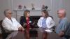
Navin Kapur, M.D., FAHA, FACC, FSCAI, director, Acute Mechanical Circulatory Support Program and executive director of The CardioVascular Center for Research and Innovation (CVCRI), Tufts Medical Center, explains how Tufts determines the level of hemodynamic support a patient needs. They use an algorithm to determine if low levels of support are needed with an intra-aortic balloon pump (IABP), or incrementally high levels with a percutaneous Impella pump, TandemHeart, extracorporeal membrane oxygenation (ECMO), or a surgically implanted ventricular assist device (VAD).


Related Cardiogenic Shock and Hemodyanmic Support Content:

VIDEO: Door-to-Unloading (DTU) Trial May Change STEMI Care

VIDEO: Tufts Uses a Hemodynamic Support Algorithm to Determine What Devices to Use

VIDEO: Hemodynamic Support Protocols at Henry Ford Hospital

VIDEO: Cardiogenic Shock Initiative Continues to Reduce Mortality by 50 Percent


VIDEO: How to Reduce Cardiogenic Shock Mortality by 50 Percent

SCAI Releases New Consensus Document on Classification Stages of Cardiogenic Shock

Cardiogenic Shock Survival Rates Improve in Three Years Since Impella FDA Approval

VIDEO: The Importance of Ventricular Unloading in AMI and Cardiogenic Shock


VIDEO: Escalation of Support and Algorithms for Cardiogenic Shock

10 Reasons Why it is Time to Learn More About Cardiogenic Shock

New Approaches to Reduce Cardiogenic Shock Mortality


Find more content on Tufts Medical Center 

Robotic Systems | January 20, 2020

This video shows the first robotic percutaneous coronary interventions (PCI) performed in Germany with the Robocath R-One robotic catheter guidance system. The first procedures were performed by Professor Michael Haude, director of Medical Clinic I at Rheinland Klinikum Neuss Lukaskrankenhaus, and his team. read more in the article First Robotic Coronary Angioplasties Performed With Robocath System in Germany.


Related Robocath Content:

Robocath Successfully Carries Out First Robotic Coronary Angioplasties in Humans

Robocath Receives $1.5 Million in Capital for Advancement of R-One Robotic System 


RSNA | January 13, 2020

DAIC/ITN Editor Dave Fornell takes a tour of some of the most innovative new medical imaging technologies displayed on the expo floor at the Radiological Society of North America (RSNA) 2019 meeting. 

Technology examples include a robotic arm to perform remote ultrasound exams, integration of artificial intelligence (AI) to speed or automate radiology workflow, holographic medical imaging display screens, a new glassless digital radiography (DR) X-ray detector, augmented reality for transesophageal echo (TEE) training, moving DR X-ray images, 3-D printed surgical implants created from a patient's CT imaging, DR X-ray tomosynthesis datasets, radiation dose management and analytics software, and new computed tomography (CT) technologies.


Find more videos and news from RSNA 2019


Wearables | January 09, 2020

The Consumer Electronic Show (CES) is the world's gathering place for consumer technologies, with more than 175,000 attendees and more than 4,400 exhibiting companies. New healthcare technologies are among the top trends at CES. This video offers a quick look at the trends specific to healthcare technology.

Artificial intelligence (AI) is one of the hottest technology trends across all product across the CES floor this year. There is also discussion by key note speakers that the internet-of-things (IOT) concept introduced at CES nearly a decade ago is now morphing into a new meaning for the interconnectivity-of-things. This can be seen in healthcare products shown here and across all types of consumer and business products. 

The device technology at CES include many examples of how integrated wearables can digitally enable healthcare. The future of healthcare will include system where consumers are continuously monitored with sensors, software and services that can pinpoint digital biomarkers — earlier warning signs that predict health events. This is the prediction of Leslie Saxon, M.D., executive director of the University of Southern California (USC) Center for Body Computing (CBC), is speaking as a panelist about digital health trends and challenges in the session “Proving the Impact of Transformative Technology.” 

Saxon is a board-certified cardiologist and digital health expert who understands how developing technologies can more accurately assess wellness and human performance among elite athletes, military personnel and patients. She explained this digital healthcare model of the future is a vast contrast to the current point-of-care model.


Cath Lab | January 09, 2020

Haval Chweich, M.D., medical director of the cardiac critical care unit (CCU) at Tufts Medical Center, and assistant professor at Tufts University School of Medicine, explains the role of intensivists on the cardiac care team. 

Chweich is an intensivist specialized in pulmonary and critical care. He interfaces with Tuft's cardiac surgeons and interventional cardiologists to care for patients as they transition after procedures into the CCU. He also plays a key role as part of the team caring for cardiogenic shock patients. 

Find VIDEOS and articles on Tufts cardiology program

Hemodynamic Support Devices | January 09, 2020

Richard Botto, CVT, RCSA, chief cardiovascular technologist, division of cardiology, cardiac cath lab, and Melissa Smith, RT, at Tufts Medical Center, Boston, explain the use of technology to remotely access data and waveforms on patients' temporary hemodynamic support system control consoles. 

Tufts Medical Center was one of the first hospitals to begin using the Abiomed Impella Connect technology, which enables remote smartphone access to Impella consoles. This allows a quick, remote check on patients using temporary hemodynamic support. The technology also is connected to a support center at Abiomed, so if a console or patient is experiencing issues out of the ordinary, techs can remote into the patient's Impella control console to take a look. Tuft's intensivists in the cardiac care unit (CCU) use the app to check on their patients' consoles without needing to walk into each room. 

Find VIDEOS and articles on Tufts cardiology program


Magnetic Resonance Imaging (MRI) | January 06, 2020

Karen Ordovas, M.D., MAS, professor of radiology and cardiology at the University of California San Francisco (UCFS) School of Medicine and a Society of Cardiac Magnetic Resonance (SCMR) board member, explains how cardiac MRI can help in women's heart disease and to better define complex congenital heart anatomy. She spoke at the 2019 Radiological Society of North America (RSNA) meeting. 

She specializes in cardiac and pulmonary imaging, and has particular expertise in using CT and MRI techniques in cardiovascular imaging and the differences of presentation in imaging between male and female cardiac patients. Ordovas is helping advance education around heart disease in women and bring great awareness of quality tools to diagnose heart disease and how heart MRI can help. She also is heavily involved in the use of heart MRIs for pregnant women, since there is no radiation,  and patients with congenital heart disease where detailed imaging of the complex anatomy is required.

The use of cardiac MRI in congenital heart disease is common in serial imaging of patients with Tetralogy of Fallot (TOF), one of the most common congenital heart diseases for which patients are referred for post-operative magnetic resonance (MR) imaging evaluation. In the past few decades, surgery has proved successful, but most patients require repeat imaging throughout their lives and MRI can offer more detailed soft tissue imaging without the use of radiation. 


Related Cardiac MRI Content:

Cardiac MRI Becoming More Widely Available Thanks to AI and Reduced Exam Times

VIDEO: Advances in Cardiac MRI Technology — Interview with James Carr, M.D.

Cardiac MRI Delivers Accurate Diagnosis for Frontline Chest Pain Evaluation

VIDEO: Dedicated Cardiac MRI Use at the Baylor Scott White Heart Hospital

Advantages and New Applications of Cardiac MRI

Will Cardiac MRI Expand?


Magnetic Resonance Imaging (MRI) | December 20, 2019

James Carr, M.D., chair of the Department of Radiology, Northwestern University, and incoming 2020 President of the Society of Cardiac Magnetic Resonance (SCMR), explains why MRI is an ideal cardiac imaging modality, at the 2019 Radiological Society of North America (RSNA) meeting.

Heart MRI offers advantages over computed tomography (CT) and echocardiography because of its excellent soft tissue delineation and its ability to offer information beyond anatomical imaging, such as perfusion, morphology and metabolism. MRI can be technically challenging and the exams requiring a long time, but recent advances have helped cur cardiac imaging times down significantly. Automation and artificial intelligence (AI) also is making post-processing and quantification mush faster, brining it closer to the time it takes to scan and post-process CT imaging.

Northwestern is was one of the early adopters of cardiovascular MRI. Carr said heart MRI was not common in regular clinical use until the past decade at some luminary centers. In 2005, Carr was given the opportunity to develop a clinical cardiac MRI program at Northwestern.
 
He said MRI scanners have improved, and now much faster than a decade ago. They are also more optimized for cardiac imaging. While heart MRI is well known in large hospital centers, Carr said it still needs to develop and expand to community hospitals and rural hospitals outside major population centers. 

Artificial intelligence is playing a significant role in cardiac MRI automation of speeding workflow and quantification. Carr said these technologies will become mainstream in the next few years. AI also will play an increasing role in risk prediction based on new image analysis algorithms in development.

For more information on cardiac MRI, visit SCMR's website www.heartmri.org.


Related Cardiac MRI Content:

Cardiac MRI Delivers Accurate Diagnosis for Frontline Chest Pain Evaluation

Cardiac MRI Becoming More Widely Available Thanks to AI and Reduced Exam Times

VIDEO: Dedicated Cardiac MRI Use at the Baylor Scott White Heart Hospital — Interview with Haojie Wang, M.D.

VIDEO: Dedicated Cardiac MRI Use at the Baylor Scott White Heart Hospital

Advantages and New Applications of Cardiac MRI

Will Cardiac MRI Expand?

VIDEO: Use of Cardiac MRI in Congenital and Women's Heart Disease — Karen Ordovas, M.D., 


Cardiovascular Ultrasound | December 20, 2019

This is the LVivo auto cardiac ejection fraction (EF) app that uses artificial intelligence (AI) from the vendor Dia, displayed at the Radiological Society Of North America (RSNA) 2019. The user opens the app in a couple seconds the AI defines to myocardial border and calculates EF for left ventricle (LV). It is shown here integrated into the GE Healthcare VScan point-of-care-ultrasound system (POCUS).

The company also partners with Konica-Minolta to supply auto EF for ultrasound images on its cardio PACS.

Read more about the system from an ASE 2019 stud

Radiation Dose Management | December 19, 2019


Mahadevappa Mahesh, Ph.D., chief of medical physicist and professor of radiology and medical physics, Johns Hopkins University, Baltimore, treasurer of the American Association of Physicists in Medicine (AAPM),a board member of the American College of Radiology (ACR), presented a late-breaking study on how medical imaging radiation dose has started to drop over the past decade. He is the co-chair of the National Council on Radiation Protection and Measures Report (NCRP), and presented the most recent NCRP data analysis at the 2019 Radiological Society of North America (RSNA) meeting.

The new NCRP 184 report covers the period between 2006 and 2016, the period of the most current CMS data. It shows a decrease of about 20 percent in the radiation dose the U.S. population receives from medical imaging, compared to the NCRP 160 that covered the period of up to 2006.

Key findings of the study include:

   • CT dose dropped about 6 percent, despite a 20 percent increase CT scans since 2006;

   • Drop of more than 50 percent for nuclear imaging scans, mainly due to fewer procedures begin performed;

   • A 15-20 percent decrease across X-ray imaging modalities.

Mahesh says this shows the impact of using "as low as reasonably achievable" (ALARA) principals, new dose guidelines outlined jointly by numerous medical societies, and dose reduction initiatives like Image Wisely, Image Gently, and the American College of radiology (ACR) Dose Index Registry.

He said there was growing concern a decade ago when the last council report was published, which show a steep increase in radiation dose. This was mainly due to a rapid increase in the use of computed tomography (CT) and other types of X-ray based and nuclear radiotracer medical imaging. This prompted the ACR top create the Image Wisely program and push for the use of more thoughtful imaging doses based on patient size, using the "as low as reasonably achievable” (ALARA) principle. While CT dose was lowered, he said the biggest decline was in nuclear imaging.


Cardiovascular Ultrasound | December 19, 2019

This is an example of an augmented reality (AR) training system for transesophageal echo (TEE) created by the simulation company CAE. Rather than just looking at an overhead screen, this system allows the user to use a HoloLens visor to see the impact their probe manipulation has on the imaging and better shows the orientation of the ultrasound probe, the 2-D ultrasound image slice and the relation to the anatomy. It was displayed at the 2019 Radiological Society Of North America (RSNA) meeting.

Read more about this technology.

Find more technology news and video from the RSNA meeting


RSNA | December 18, 2019

DAIC Editor Dave Fornell and Imaging Technology News (ITN) Consulting Editor Greg Freiherr offer a post-game report on the trends and technologies they saw on the expo floor of 2019 Radiological Society of North America (RSNA) annual meeting. This includes artificial intelligence (AI), augmented reality, holographic imaging, cybersecurity, and advances in digital radiography (DR) with a glassless detector plate, X-ray tomosynthesis, dual-energy X-ray and dynamic DR imaging. 

Find RSNA news and other videos

Nuclear Imaging | November 07, 2019

Rob Beanlands, M.D., FASNC, 2019 American Society of Nuclear Cardiology (ASNC) president, shares a couple trends he sees in cardiac nuclear imaging. He is the Vered Chair and division head of cardiology and director of the National Cardiac PET Centre at the University of Ottawa Heart Institute, Canada.

He said overall trends he sees in nuclear cardiology include the use of better myocardial reserve quantification so it is clear whether revascularization would help patients. Beanlands also said there is increasing interest in positron emission tomography (PET) imaging because of its superior image quality and increasing access to PET radiotracers. New tracers on the horizon will also increase the image quality and flexibility of PET to accommodate exercise stress.

He spoke on artificial intelligence (AI) development for medical imaging in sessions at the 2019 American Society of Nuclear Cardiology (ASNC) annual meeting. 

Artificial Intelligence | November 07, 2019

Piotr J. Slomka, Ph.D., FACC, research scientist in the Artificial Intelligence in Medicine Program, Department of Medicine at Cedars-Sinai, and professor of medicine in-residence of the David Geffen School of Medicine, UCLA. He explains how his team at Cedars-Sinai is working on intelligent patient risk prediction algorithms that will automatically extract information from medical imaging. He spoke on artificial intelligence (AI) development for medical imaging in sessions at the 2019 American Society of Nuclear Cardiology (ASNC) annual meeting. 

Find more articles and video on AI


Nuclear Imaging | November 06, 2019

Robert C. Hendel, M.D., explains some of the new cardiac radiotracers in the pipeline that were discussed in sessions at the American Society of Nuclear Cardiology (ASNC) 2019 meeting. Hendel is a professor of medicine and radiology and the Sidney W. And Marilyn S. Lassen Chair in Cardiovascular Medicine, Tulane University. He also serves as the chief of the section of cardiology and director of the Tulane University Heart and Vascular Institute. 

He outlined three new radiotracers that may impact cardiac imaging:

   • Flurpiridaz F-18 PET perfusion agent that offers high quality images and can be used with exercise stress;

   • MIBG imaging to help better assess heart failure classes and if patients may need an ICD;

   • New F-18 agents to image cardiac amyloidosis; and

   • Nuclear agents to enable annexin imaging, allowing direct vsualization of myocardial apoptotic cells.


PET-CT | October 30, 2019

Rupa Sanghani, M.D., FASNC, associate professor, Rush Medical College, director of nuclear cardiology and stress laboratory, Rush University Medical Center, and associate director, Rush Heart Center for Women, explains how to create a high-volume cardiac positron emission tomography (PET) imaging program. She spoke on this topic at the 2019 meeting of the American Society Nuclear Cardiology (ASNC) and led a tour for attendees of the PET-CT system at Rush, which was located close to the conference. 

Sanghani said the advantages of PET myocardial perfusion imaging include faster exam times and allowing additional information from coronary reserve flow assessments to better understand if revascularization will help a patient's heart recover. The 16-slice CT scanner is used not only to attenuate the PET images, but to perform a CT calcium scoring exam to assess the patient's risk for future cardiovascular events. The Rubidium-82 radiotracer used for PET exams only has a 75 second half life, so it can help increase the number of exams a center is able to perform each day. At higher volume centers, PET is optimized to handle all the patients who require pharmacological stress exams. 

In the video, Sanghani  outlines what Rush did to design its room, covers basics on training, what to look for in a scanner and other considerations when creating a PET program.

Read the article How to Start a Cardiac PET-CT Imaging Program, which includes more information from the Rush PET program.
 

Find more coverage of the ASNC

Find more news on nuclear imaging


Cardiac Diagnostics | October 29, 2019

Clyde Yancy, M.D., MSc, cardiology chief and vice dean for diversity and inclusion at Northwestern University, Feinberg School of Medicine, was a keynote speaker at the 2019 American Society of Nuclear Cardiology (ASNC) annual meeting. He said the traditional biases of seeing a patient and automatically making clinical assumptions because they are a certain race or gender are obsolete. For example, he said not all black patients have hypertension. Yancy added that genetics, especially with racial intermarriage over the past several generations, no longer predisposes patients to what is typically assumed for certain ethnic or racial backgrounds.

Yancy also said new research is showing how diet plays a major role in patient health and disease progression for things previously thought to be based on genetics. This includes the people who live in "food deserts" in urban areas where there are no sources of fresh food and vegetables, so they consume large amounts of packaged and processed foods that contain high levels of salt, phosphates and preservatives. He said these chemicals and diet may be the root cause of hypertension and diabetes in black populations in low income areas, rather than genetics as previously thought.


Related Content: 

VIDEO: Reducing Hypertension Among African-Americans — Interview with Kim Allan Williams, Sr., M.D.

VIDEO: Use of Plant-Based Diet to Reduce Cardiovascular Disease Risk — Interview with Kim Allan Williams, Sr., M.D.

VIDEO: New PLATINUM Diversity Data Shows Early DAPT Cessation OK in Minorities With New Generation Stent — Interview with Roxana Mehran, M.D., 


Radial Access | October 22, 2019

Sunil Rao, M.D., chief of cardiology, Durham VA Health System and a professor at Duke University, and Prashant Kaul, M.D., director of the cath lab, Piedmont, Atlanta, discuss trends in radial access at the 2019 Transcatheter Cardiovascular Therapeutics (TCT) annual meeting. They discuss how radial access adoption has grown rapidly in the past few years and now makes up between 40-50 percent of percutaneous coronary intervention (PCI) procedural volume in the United States. They also discuss recent clinical trial data and the new concept of using distal radial access. 


Related Radial Access Content:

SCAI Updates Expert Consensus Statement on Best Practices for Transradial Access

Transradial Access Celebrates 25 Years

VIDEO: History of Radial Artery Access — Interview with Ferdinand Kiemeneij, M.D.

Incorporating Distal Radial Artery Access Into Clinical Practice

VIDEO: New Frontiers in Radial Access — Interview with Mladen I. Vidovich, M.D.

Find more news and on transradial access technique and technology

Radial Access Adoption in the United States


Vascular Closure Devices | October 17, 2019

Ashish Pershad, M.D., chief of interventional cardiology, Banner University Medical Center, Phoenix, explains the trend of using closure devices to seal larger vascular access sites from the use of TAVR, EVAR, TMVR and hemodynamic support devices at the 2019 Transcatheter Cardiovascular Therapeutics (TCT) meeting. He was one of the moderators on a session on this topic at TCT 2019.


Related Large Bore Vascular Closure Device Content:

VIDEO: How to Achieve Hemostasis With Large Bore Device Access — Interview with Philippe Genereux, M.D.

First-in-Human Results Show Early Bird Device Effective in Early Detection of Internal Bleeding

Advances and Trends in Vascular Closure Devices

Manta Large-Bore Vascular Closure Device Cleared by the FDA

PerQseal Large Bore Closure Device Launches in Europe

Teleflex Acquires Essential Medical


Related Content With Dr. Pershad:

National Coverage Determination Will Make TAVR Available to More Patients at More Centers

VIDEO: Comparison Between Watchman vs. Amulet LAA Occluders
 

Antiplatelet and Anticoagulation Therapies | October 17, 2019

Roxana Mehran, M.D., FACC, FACP, FCCP, FESC, FAHA, FSCAI, professor of medicine and director of interventional cardiovascular research and clinical trials at the Zena and Michael A. Wiener Cardiovascular Institute at Mount Sinai School of Medicine, explains the use of short dual antiplatelet therapy (DAPT) in minorities from the PLATINUM Diversity Trial. The first trial data was released in 2017, and she presented new data from the study at the 2019 Transcatheter Cardiovascular Therapeutics (TCT) meeting. 

The study looked at use of the Promus Element Plus Post-Approval Study and the clinical impact of dual-antiplatelet therapy (DAPT) cessation within 12 months of drug-eluting stent implantation in caucasians and minorities. It is one of the first large trials to stress the importance of diversity in clinical trials, which tent to reflect a population of older white men.


Find more news and videos from TCT 2019

Hemodynamic Support Devices | October 16, 2019

Jeffrey J. Popma, M.D., director of interventional cardiology clinical services at Beth Israel Deaconess Medical Center, professor of medicine at Harvard Medical School, explains the results of the PROTECT II and the new PROTECT III Study at the 2019 Transcatheter Cardiovascular Therapeutics (TCT) meeting. PROTECT III is the follow up to PROTECT II RCT and the largest-ever FDA study of hemodynamically supported, high-risk PCI patients. 

He discusses the PROTECT II and PROTECT III studies, and real-life patient data from the Impella IQ Database. 


Find more news and videos from TCT 2019

Structural Heart | October 16, 2019

Vivian Ng, M.D., assistant professor of medicine and an interventional cardiologist at the NewYork-Presbyterian/Columbia University Medical Center Structural Heart and Valve Center, helped organize the first Women in Structural Heart (WISH) event at the 2019 Transcatheter Cardiovascular Therapeutics (TCT) meeting. The evening session was standing room only and highlighted structural heart case presentations and discussion panels made up of all women. The session panelists and presenters were a whose-who of well known women in cardiology. The event was organized as a way to break the glass ceiling in the subspecialty of interventional cardiology, where women make up less than 5 percent of the operators.


Previous Video Interviews With Speakers and Panelists Involved in the WISH Session:

VIDEO: The Importance of the Neo-LVOT in Transcatheter Mitral Valve Replacement — Interview with Dee Dee Wang, M.D.

VIDEO: The Value of the Cardiovascular Service Line — Interview with Linda Gillam, M.D.,

VIDEO: Can We Live in 3-D Echo? — Interview with Lissa Sugeng, M.D.

VIDEO: Tricuspid Valve Imaging and Interventions Developing Hand-in-hand — Interview with Rebecca Hahn, M.D.

VIDEO: Strategies to Avoid Acute Kidney Injury Caused by Cath Lab Contrast — Interview with Roxana Mehran, M.D.,


Find more news and videos from TCT 2019

Antiplatelet and Anticoagulation Therapies | October 11, 2019

Ajay J. Kirtane, M.D., associate professor of medicine at Columbia University Irving Medical Center and director of the cardiac catheterization laboratories at NewYork-Presbyterian (NYP) Hospital, shares the findings of the late-breaking EVOLVE Short DAPT study presented as a late-breaking trial at the 2019 Transcatheter Cardiovascular Therapeutics (TCT) meeting. It evaluated the safety of early dual antiplatelet therapy (DAPT) discontinuation in patients with high bleeding risk treated with the a Boston Scientific Synergy bioabsorbable polymer coated drug-eluting stent (DES).

With the advancement of new DES technologies using thinner struts and new types of drug-carrier polymer techniques, the risk of late-stent thrombosis has been greatly reduced, meaning there is less need for long-term DAPT. For patients who are at high risk for bleeding, who have ulcers or other types of gastrointestinal (GI) bleeding, or those going into surgery, it would be beneficial to reduce the time period for DAPT, and several late-breaking trials examined this at TCT 2019.
 

Here are the other late-breaking DAPT studies:


Find more news and videos from TCT 2019

Antiplatelet and Anticoagulation Therapies | October 10, 2019

Roxana Mehran, M.D., FACC, FACP, FCCP, FESC, FAHA, FSCAI, professor of medicine and director of interventional cardiovascular research and clinical trials at the Zena and Michael A. Wiener Cardiovascular Institute at Mount Sinai School of Medicine, explains insights from the Ticagrelor With Asprin or Alone in High-Risk patients after Coronary Intervention (TWILGHT) Trial. She presented this late-breaking study at the 2019 Transcatheter Cardiovascular Therapeutics (TCT) meeting. Here is a link to the article on the TWILIGHT Trial.


Related Short DAPT Content:

VIDEO: Overview of Short DAPT in High-risk Bleeding Patients Who Receive Stents — Interview with AHA President Robert Harrington, M.D. at TCT 2019 

Onyx ONE: A Randomized Trial of a Durable-Polymer Drug-Eluting Stent vs a Polymer-Free Drug-Coated Stent in Patients at High Risk of Bleeding Treated With 1-Month DAPT - TCT 2019 late-breaker

IDEAL-LM: A Randomized Trial of a Bioabsorbable Polymer DES With 4-Month DAPT vs a Durable Polymer DES With 12-Month DAPT in Patients With Left Main Coronary Artery Disease - TCT 2019 late-breaker 

EVOLVE Short DAPT: A Single Arm Study of 3-Month DAPT in Patients at High Bleeding Risk Treated a Bioabsorbable Polymer-Based Everolimus-Eluting Stent - TCT 2019 late-breaker 

New Directions and Trends in Coronary Metallic Stents

Questions Remain on DAPT Prolongation


Find more TCT late-breaking news and video

Heart Valve Technology | October 09, 2019

Torsten Vahl, M.D., director of experimental and translational research, Structural Heart and Valve Center and at the Center for Interventional Vascular Therapy at NewYork-Presbyterian Hospital/Columbia University Medical Center, explains the current state of transcatheter aortic valve replacement (TAVR) following the U.S. Food and Drug Administration (FDA) clearance of TAVR in all surgical risk patient categories. Europe also cleared the use of the Sapien 3 device for low risk patients in November 2019.

He spoke on TAVR at the Transcatheter Cardiovascular Therapeutics (TCT) 2019 meeting. In August 2019, the FDA cleared the use of TAVR in all surgical risk classes of patients using the Edwards Sapien 3 and Medtronic CoreValve devices. This means the heart team made up of a cardiac surgeon, interventional cardiologist and cardiac imaging specialist can determine if surgical aortic valve replacement (SAVR) or TAVR should be used. It is widely expected TAVR use will rapidly rise in the next five years to take over 50 to 70 percent of the aortic valve replacement procedural volume. It is expected surgical volumes will drop to about 20 percent of patients who do not qualify for TAVR because of anatomic issues or other variables that make SAVR a safer option.


Related Transcatheter Valve Content:

FDA Approves TAVR for Low-risk Patients Creates A Paradigm Shift in Cardiology

Edwards Sapien 3 TAVI Granted European Approval to Treat Low-risk Patients

VIDEO: Tracking Transcatheter Valve Outcomes in the STS-ACC TVT Registry — John Carroll, M.D.

VIDEO: Transcatheter Mitral Valve Replacement Planning — Interview with Joao Cavalcante, M.D.

VIDEO: Overview of University of Colorado Structural Heart Program — Interview with John Carroll, M.D., Robert Quaife, M.D., and James Chen, Ph.D.

Advances in Transcatheter Tricuspid Valve Technologies


VIDEO: Update of Mitral Valve Repair and Replacement Technologies at TCT 2017 — Interview with Ted Feldman, M.D.,

TAVR Operator and Hospital Requirements Outlined in 2018 AATS/ACC/SCAI/STS Expert Consensus

VIDEO: Tricuspid Device Clinical Trial Overview — Interview with Ori Ben-Yehuda, M.D.

Recent Advances in Transcatheter Valve Technology


VIDEO: Tricuspid Valve Imaging and Interventions Developing Hand-in-hand at ASE 2018— Interview with Rebecca Hahn, M.D.

Hospital Consolidation May Increase Access to TAVR, New Cardiac Technologies

VIDEO: TAVR for Degenerated Surgical Valves - 2017 Update— Valve-in-Valve TAVR Procedures — Interview with Sammy Elmariah, M.D.

How to Perform Transcaval TAVR Access


VIDEO: The Essentials of CT Transcatheter Valve Imaging — Interview with Jonathon Leipsic, M.D.

Advances in Heart Valve Technology in 2017

VIDEO: TAVR For Asymptomatic Severe Aortic Stenosis — Interview with Philippe Genereux, M.D.

VIDEO: Conscious Sedation for TAVR Procedures — Interview with Mario Goessl, M.D.


VIDEO: Overview of the Henry Ford Hospital Structural Heart Program

Advances and Future Directions for Transcatheter Valves

Transcatheter Mitral Valve Replacement Devices in Development


Antiplatelet and Anticoagulation Therapies | October 08, 2019

American Heart Association President Robert Harrington, M.D., interventional cardiologist and the Arthur L. Bloomfield Professor of Medicine and chairman of the Department of Medicine at Stanford University, explains the reasons for shorter duration dual-antiplatelet therapy (DAPT) in high-risk bleeding patients. Newer-generation drug-eluting stents are designed for faster vessel healing and endothelialization, so the time required for DAPT can be reduced from 12 months to a shorter time period. At the 2019 Transcatheter Cardiovascular Therapeutics (TCT) meeting there were several late-breaking trials that looked at shorter duration DAPT or use of mono-therapy with ticagrelor. 

Related Short DAPT Content:

TWILIGHT: A Randomized Trial of Ticagrelor Monotherapy vs Ticagrelor Plus Aspirin Beginning at 3 Months in High-risk Patients Undergoing PCI - TCT 2019 late-breaker

Onyx ONE: A Randomized Trial of a Durable-Polymer Drug-Eluting Stent vs a Polymer-Free Drug-Coated Stent in Patients at High Risk of Bleeding Treated With 1-Month DAPT  - TCT 2019 late-breaker

IDEAL-LM: A Randomized Trial of a Bioabsorbable Polymer DES With 4-Month DAPT vs a Durable Polymer DES With 12-Month DAPT in Patients With Left Main Coronary Artery Disease - TCT 2019 late-breaker

EVOLVE Short DAPT: A Single Arm Study of 3-Month DAPT in Patients at High Bleeding Risk Treated a Bioabsorbable Polymer-Based Everolimus-Eluting Stent  - TCT 2019 late-breaker


Cardiogenic Shock | October 08, 2019

William O’Neill, M.D., medical director of the Center for Structural Heart Disease at Henry Ford Hospital, Detroit, explains data on first 250 patients in the National Cardiogenic Shock Initiative Study (NCSI) and new escalation protocols during the Transcatheter Cardiovascular Therapeutics (TCT) 2019 meeting. By implementing a protocol of using Impella hemodynamic support prior to percutaneous coronary intervention (PCI), the 80 centers involved in the initiative have been able to increase survival by 50 percent.


Related Cardiogenic Shock Content:

VIDEO: How to Reduce Cardiogenic Shock Mortality by 50 Percent — Interview with Babar Basir, D.O.

SCAI Releases New Consensus Document on Classification Stages of Cardiogenic Shock

VIDEO: The Importance of Ventricular Unloading in AMI and Cardiogenic Shock — Interview with Nevin Kapur, M.D.

10 Reasons Why it is Time to Learn More About Cardiogenic Shock — by Emmanouil S. Brilakis, M.D.

New Approaches to Reduce Cardiogenic Shock Mortality

VIDEO: Overview of the National Cardiogenic Shock Initiative — Interview with William O’Neill, M.D.


Find more TCT late-breaking news and video


TCT | October 04, 2019

Chandan Devireddy, M.D., offers insights about what he saw as the top take aways from the 2019 Transcatheter Cardiovascular Therapeutics (TCT) annual meeting. He is cath lab director at Emory University Hospital Midtown, associate professor of medicine of interventional cardiology at Emory University, and chairperson for the Society of Cardiovascular Angiography and Interventions (SCAI) Communications Committee. 

Devireddy said his key take aways from TCT 2019 were:

   • The poor results of the Boston Scientific Acurate Neo self-explaining TAVR system compared to the current devices on the U.S. market;

   • Results of the Abbott Portico TAVR system;

   • Levant Trial shows no increased mortality due to use of a paclitaxel-coated balloon; and

   • CT study of TAVR valve leaflet thrombosis.


Find more TCT late-breaking news and video


University of Colorado Hospital | October 02, 2019

Interview with John Carroll, M.D., director of interventional cardiology, Robert Quaife, M.D., director of advanced cardiac imaging, and James Chen, Ph.D., associate professor of medicine and director of the 3-D imaging lab at the Cardiac and Vascular Center at the University of Colorado Hospital. They discuss how the structural heart program was created and how they invested in advanced imaging to grow into one of the most advanced programs in the country. They explain how the program now incorporates transcatheter aortic valve replacement (TAVR), transcatheter mitral valve repair, transcatheter mitral valve replacement (TMVR), left atrial appendage (LAA) occlusion and transcatheter closure of holes in the heart. 

The heart team in this video stressed the need for advanced imaging to plan and guide the procedures. They explain how the center developed its own 3-D imaging software and worked with Philips healthcare to commercialize some of the technologies, including the EchoNavigator system used to fuse live angiography with live transesophageal echo (TEE).


Related University of Colorado Hospital Content:

Highlighting Innovation at the University of Colorado Hospital Cardiology Program

VIDEO: Evolution of Transcatheter Mitral Valve Repair at the University of Colorado — Interview with John Carroll, M.D., and Robert Quaife, M.D.

VIDEO: The Role of Advanced Imaging in Structural Heart Interventions — Interview with Robert Quaife, M.D.

VIDEO: Advice For Hospitals Starting a Structural Heart Program — Interview with John Carroll, M.D.

VIDEO: The Evolution of Complex PCI at University of Colorado — Interview with John Messenger, M.D., and Kevin Rogers, M.D.

VIDEO: Developing New Cath Lab Technologies With Real-time Collaboration Between Industry, Doctors

360 View of the TEE Echo Workstation During a MitraClip Procedure

VIDEO: Walk Through of a Hybrid Cath Lab at the University of Colorado Hospital

VIDEO: Cath Lab Walk Through at the University of Colorado Hospital

VIDEO: The Cardiac Surgeon Perspective on Transcatheter Mitral Valve Repair — Interview with Joe Cleveland, M.D.

VIDEO: An Overview of PFO Closure to Treat Cryptogenic Stroke — Interview with Karen Orjuela, M.D.,


(This video was originally posted in May 2019 and was updated Oct. 2, 2019)


Cardiovascular Business | September 30, 2019

A discussion with Ruth Fisher, MBA, vice president of the Henry Ford Hospital structural heart program, and Janet Wyman, NP, program manager. They explain how Henry Ford has been able to build its program and work with hospitals throughout Michigan, including those with their own transcatheter aortic valve replacement (TAVR) programs. They said building relationships with referral centers as partners and ensuring the patients go back to their local physicians for followups and regular care is key. 

Watch the related VIDEO: Overview of the Henry Ford Hospital Structural Heart Program.

(Editor's note - this article was originally published in April 2019 and was revised Oct. 1, 2019)

Additional articles and videos on Henry Ford Hospital 


Heart Valve Technology | September 30, 2019

Marvin Eng, M.D., structural fellowship director at Henry Ford Health System, and William O'Neill, M.D., director of the Henry Ford Hospital structural heart program, explain the mitral valve repair program at Henry Ford. 

The hospital performs a large number of transcatheter mitral valve repairs, including MitraClip and with its involvement in trials for investigational device technologies. 

Watch the VIDEO: Overview of the Henry Ford Hospital Structural Heart Program

Find more news and video from Henry Ford Hospital

(Editor's note - this article was originally published in March 2019 and was revised Sept. 30, 2019)


Structural Heart | September 30, 2019

A discussion with William O’Neill, M.D., director of the Henry Ford structural heart program, Ruth Fisher, MBA, vice president of the program, and Janet Wyman, NP, program manager. Henry Ford Hospital has built a large, high-volume structural heart program that includes use of cutting edge new technologies and devices. 

Watch the related VIDEO: How to Get Referral Patients Into a Structural Heart Program — Henry Ford Hospital shares insights from its program.
 

Additional articles and videos on Henry Ford Hospital 
 

(Editor's note - this video was originally published in September 2018 and was revised September 2019)

Cath Lab | September 28, 2019

A discussion with Nicolas Bevins, Ph.D., vice chair, physics and research, and Jessica Harrington, RCIS. They explain the use of shields, technique and use of newer angiography technologies to reduce X-ray radiation dose in the cardiac cath labs at Henry Ford Hospital, Detroit.

Watch the VIDEO: Technologies and Techniques to Reduce Radiation Dose in the Cardiac Cath Lab — Interview with Akshay Khandelwal, M.D., director of medical operations at the Henry Ford Heart and Vascular Institute

Additional articles and videos on Henry Ford Hospital 


For more on how to reduce dose in the cath lan, read these related articles:

Cardiology Societies Call for Better Radiation Dose Tracking

Defining the Cath Lab Workplace Radiation Safety Hazard

Dose-Lowering Practices for Cath Lab Angiography

5 Technologies to Reduce Cath Lab Radiation Exposure

VIDEO: Heart Surgeon Shares Effects of Fluoroscopic Radiation Exposure

Helping Interventional Cardiologists Reduce Exposure to Ionizing Radiation

14 Ways to Reduce Radiation Exposure in the Cath Lab


(Editor's note - this video was originally published in September 2018 and was revised September 2019)

September 26, 2019

Clifford Robinson, M.D., associate professor of radiation oncology, chief of the SBRT service, director of clinical trials, Washington University, St. Louis, Washington University School of Medicine in St. Louis, explains the longer term results of cardiac radiotherapy ablation to treat ventricular tachycardia. Image-guided cardiac radioablation dramatically reduces episodes of fast, abnormal heartbeats for more than two years in the ENCORE-VT trial.

In collaboration with Phillip Cuculich, M.D., associate professor of cardiology and radiation oncology at the Washington University School of Medicine in St. Louis, Robinson and his team developed a noninvasive, outpatient procedure for treating VT called EP-guided noninvasive cardiac radioablation (ENCORE-VT). This novel therapy fuses electrical (ECGs) and imaging (computed tomography, magnetic resonance imaging, positron emission tomography) data to pinpoint the scar tissue in the patient’s heart responsible for the arrhythmias, then targets it with a single dose of stereotactic body radiation therapy (SBRT), a type of high-dose radiation most commonly used to treat patients with cancer. ENCORE-VT requires no general anesthesia and allows patients to go home immediately after treatment. 

The results of the ENCORE-VT study were presented at ASTRO 2019. The results were also presented as a late-breaking trial at the Heart Rhythm Society (HRS) 2020 virtual meeting.

Read the article Noninvasive Radioablation Offers Long-term Benefits to High-risk Heart Arrhythmia Patients.


Related Radiotherapy for Cardiac Ablation Content:

Radiation Therapy to Treat Heart Arrhythmias is a Technology to Watch

New Technologies to Improve Atrial Fibrillation Ablation

New Targets for Radiation Therapy Include Cardiac AF Ablations and Renal Denervation

VIDEO: Using Radiosurgery for Atrial Fibrillation Cardiac Ablation

CyberHeart to Develop Noninvasive Cardiac Ablation Technique Using Accuray Robotic System

Radiation Therapy to Treat Heart Arrhythmias is a Technology to Watch

VIDEO: Use of Radiotherapy to Noninvasively Ablate Ventricular Tachycardia — Interview with Pierre Qian, MBBS

Varian Acquires CyberHeart Cardiac Radio-ablation Technology

Noninvasive Radioablation Offers Long-term Benefits to High-risk Heart Arrhythmia Patients

Find more radiation oncology news


Hemodynamic Support Devices | September 22, 2019

There was a 77 percent increase in survival in cardiogenic shock patients treated using a new protocol in the National Cardiogenic Shock Initiative. The data was presented at the 2018 Transcatheter Cardiovascular Therapeutics (TCT) meeting. The details are discussed here with Babar Basir, D.O., and William O'Neill, M.D., from Henry Ford Hospital

Watch the related VIDEO: Overview of the National Cardiogenic Shock Initiative

Watch the related VIDEO: Support Protocols at Henry Ford Hospital

Additional articles and videos on Henry Ford Hospital 

(Editor's note - this video was originally published in September 2018 and was revised September 2019)

Chronic Total Occlusion (CTO) | September 28, 2019

A discussion with Khaldoon Alaswad, M.D., director, cardiac catheterization lab, Henry Ford Hospital, on treating chronic total occlusions (CTOs) and other complex PCI cases. Watch the VIDEO: Treating Chronic Total Occlusions with Bill Lombardi, M.D., director of complex coronary artery interventions at the University of Washington. Read the article "How to Tackle Coronary CTOs."

Additional articles and videos on Henry Ford Hospital 


Related Chronic Total Occlusion Content: 

VIDEO: New Technology to Treat Chronic Total Occlusions (CTOs), an interview with Farouc Jaffer, M.D., Ph.D.

When to Consider Revascularization of Coronary Chronic Total Occlusions

VIDEO: Treating Chronic Total Occlusions, an interview with Bill Lombardi, M.D.

VIDEO: The Evolution of Complex PCI at University of Colorado  — Interview with John Messenger, M.D., and Kevin Rogers, M.D.

How to Tackle Coronary CTOs


(Editor's note - this video was originally published in September 2018 and was revised September 2019)

Hemodynamic Support Devices | September 12, 2019

A discussion with William O'Neill, M.D., director of the structural heart program, Henry Ford Hospital, and Michele Voeltz, M.D., fellowship program director, interventional cardiology, explaining their process for selecting various levels of hemodynamic support.

Henry Ford Hospital also helped spearhead the Detroit Cardiogenic Shock Initiative that morphed into the National Cardiogenic Shock Initiative using these same protocols. Watch an interview with O'Neill explaining the national initiative.

Additional articles and videos on Henry Ford Hospital 

(Editor's note - this video was originally published in September 2018 and was revised September 2019)


Chronic Total Occlusion (CTO) | August 23, 2019

Interview with Kevin Rogers, M.D., director of vascular medicine at the University of Colorado Hospital. He explains what wires he uses for chronic total occlusions (CTOs). The university of Colorado is a high-volume CTO center. Read more about the Innovation at the University of Colorado Hospital Cardiology Program.


Related Chronic Total Occlusion Content: 

VIDEO: How to Treat CTOs and Complex PCI Cases — Interview with Khaldoon Alaswad, M.D.

Recent Advances in Interventional Guidewire Technology

VIDEO: New Technology to Treat Chronic Total Occlusions (CTOs), an interview with Farouc Jaffer, M.D., Ph.D.

When to Consider Revascularization of Coronary Chronic Total Occlusions

VIDEO: Treating Chronic Total Occlusions, an interview with Bill Lombardi, M.D.

VIDEO: The Evolution of Complex PCI at University of Colorado  — Interview with John Messenger, M.D., and Kevin Rogers, M.D.

How to Tackle Coronary CTOs

Advanced Visualization | August 09, 2019

An example of Siemens' photo-realistic Cinematic image reconstruction. This image is from a CTA exam from a Siemens Force CT scanner. 

Vendors who offer this realistic type of CT image rendering say it is not used for diagnostics. However, the technology can be helpful when explaining things to the patient and their family, educating physicians and staff, and for surgeons, since it offers a realistic view of the anatomy that is easier for most people to understand who are not familiar with cardiac anatomy as it appears in traditional CT multiplanar reconstruction (MPR) images. 

This example of software was demonstrated on the expo floor at the 2019 Society of Cardiovascular Computed Tomography (SCCT) meeting. 


Find more news and videos from the SCCT meeting

CT Angiography (CTA) | August 08, 2019

This is a quick video example of a cardiac computed tomography (CT) exam showing a Medtronic CoreValve transcatheter aortic valve replacement (TAVR) device implanted. The image was reconstructed using Canon Medical’s Global Illumination photo-realistic rendering advanced visualization post-processing software. Vendors who offer this realistic type of CT image rendering say it is not used for diagnostics. However, the technology can be helpful when explaining things to the patient and their family, educating physicians and staff, and for surgeons, since it offers a realistic view of the anatomy that is easier for most people to understand who are not familiar with cardiac anatomy as it appears in traditional CT multiplanar reconstruction (MPR) images. 

This example of software was demonstrated on the expo floor at the 2019 Society of Cardiovascular Computed Tomography (SCCT) meeting. 


Subscribe Now