Videos

VIDEO: One on One with Hal Wolf, FHIMSS, HIMSS President and CEO

HIMSS | March 05, 2024

Industry trade shows and conferences seem to be making their comeback in 2024. And the Healthcare Information and Management Systems Society (HIMSS) Global Conference and Exhibition seems particularly poised to deliver the best of the best when it comes to digital transformation in both the delivery of healthcare, but also the delivery of a quality experience for those in this demanding, rapidly evolving industry. This month in our ongoing One on One series with industry leaders, we are talking with Hal Wolf, FHIMSS, president and CEO of HIMSS. He offered insights on the society’s new partnership with Informa Markets, key topics being covered at HIMSS24, AI’s impact on the industry, and his thoughts on healthcare sustainability.

Find more HIMSS24 conference coverage here

A New Partnership for Growth

Last August, Informa Markets and HIMSS announced a landmark partnership to propel the growth and evolution of the HIMSS Global Health Conference and Exhibition, recognized as the most influential healthcare technology event of the year, and in North America. It draws 40,000 health professionals, tech leaders, providers and governmental organizations from across the globe. Informa Markets, the world’s largest exhibition organizer, took on management of the HIMSS Exhibition, while HIMSS continues to oversee developing expert content and programming.

Exciting New Features at HIMSS24

At HIMSS2024, with this new collaboration comes new features, including:

Related content:

Find more HIMSS24 conference coverage here

HIMSS Launches Modernized Infrastructure Adoption Model to Support Global Digital Health Transformation

Top Public Policy Experts at HIMSS24 to Address Global AI Landscape and Digital Transformation in Healthcare

VIDEO: Using Maturity Models to Measure Digital Health

VIDEO: Moving Digital Transformation Forward in Healthcare

VIDEO: Key Components to Creating and Implementing AI and Digital Transformation Solutions

VIDEO: The Benefits and Pitfalls of Artificial Intelligence in Healthcare

VIDEO: A Look at Cybersecurity and How Healthcare is at Risk

Recent Video

CT Angiography (CTA) | August 08, 2019

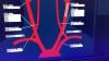
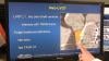
This is an example of an automated calcium scoring software to speed review of coronary artery calcium (CAC) scoring cardiac computed tomography (CT) scans. This advanced visualization software from Ziosoft uses artificial intelligence to segment the coronary vessels, identify valves and the aorta and then color code tag the calcium deposits and quantify the amount of calcified plaque in each vessel. It tallies the score into a table and computes an overall Agatston risk score. This risk score correlates to that patient's risk for a heart attack in the future. The software notes calcium in the heart outside the coronaries in valve leaflets and the aorta, but excludes this data. This type of automation is now offered by most advanced visualization and CT system vendors. This automation can save a large amount of post-processing time and make it easier for hospitals to offer low-cost CAC CT screening programs. 

CAC scans can be used to determine if a patient needs to go on statin therapy. An Agatston score of zero means the patient has no risk of coronary disease. 

Calcium in arteries is a marker for damage caused by vessel wall inflammation from atherosclerosis. Calcium can form from previously ruptured necrotic, lipid core plaques, also referred to as vulnerable plaques. These are the types of plaque responsible for heart attacks. When the core of these plaques rupture, the blood reacts to the exposed core similar to a wound and begins to clot, forming a thrombus in the vessel, which can block the blood flow. When the vessel heals over time it calcifies, leaving behind an easily identifiable marker on CT imaging. 

This example of software was demonstrated on the expo floor at the 2019 Society of Cardiovascular Computed Tomography (SCCT) meeting. 


Related CT Calcium Scorining Content:

VIDEO: The History of CT Calcium Scoring — Interview with Arthur Agatston, M.D.

VIDEO: New Cholesterol Guidelines Support CT Calcium Scoring for Risk Assessment — Interview with Matthew Budoff, M.D.

CT Calcium Scoring Becoming a Key Risk Factor Assessment

ACC and AHA Release Updated Cholesterol Guidelines for 2018

VIDEO: CT Calcium Scoring to Screen For Who Should Take Statins — Interview with Matthew Budoff, M.D.


Computed Tomography (CT) | August 07, 2019

This is an example of in-stent restenosis shown using spectral CT imaging. This example was demonstrated by Philips Healthcare at the 2019 meeting of the Society Of Cardiovascular Computed Tomography (SCCT). It can sometimes be difficult to determine if showing inside a stent of CT is artifact or real. Spectral CT can definitely help make a diagnosis. This example shows Philips’ Effective Z visualization, where the image is created based on the atomic number of the elements present in the image. After removal of iodine from the image, it shows the shadow in the stent has the same atomic composition as the vessel intima.  

This example is from a Philips IQon dual-energy CT scanner. This system has spectral imaging always on without any need to change protocols. This allows the spectral to be used when needed for additional information without need to rescan a patient.

Read more on this CT system.

VIDEO: Applications of Spectral CT — Interview with Suhny Abbara, M.D.


Cardiac Imaging | July 30, 2019

Nate Bachman, graduate research assistant in the Human Cardiovascular Physiology Lab of the Dept. of Health and Exercise Science at Colorado State University, describes how he and fellow researchers used multiple types of cardiac imaging to evaluate the cardiovascular health of athletes who compete in endurance events lasting six hours or more, and what the results may suggest for future screening.

EP Lab | July 26, 2019

Mark Ibrahim, M.D., FACC, assistant professor of medicine and radiology, associate program director, advanced cardiac imaging fellowship, University of Utah, explains what radiologists and cardiologists need to know what is needed from CT imaging prior to ablation procedures for atrial fibrillation (AF) and ventricular fibrillation (VF). He spoke at a joint session of the Heart Rhythm Society (HRS) and the Society of Cardiovascular Computed Tomography (SCCT) at the 2019 SCCT meeting. 


Find more SCCT news and videos

CT Angiography (CTA) | July 26, 2019

Andrew Choi, M.D., FACC, FSCCT, co-director, cardiac CT and MRI, assistant professor of medicine and radiology, George Washington University, Division of Cardiology, Washington, D.C., explains the role of social media to extend the reach of cardiovascular research from peer review journals. He spoke on this topic in multiple sessions at the Society of Cardiovascular Computed Tomography (SCCT) 2019 meeting.


Find more SCCT news and videos


EP Lab | July 25, 2019
Heart Valve Technology | July 24, 2019

Joao Cavalcante, M.D., FSCCT, director of structural heart CT and cardiac MRI, Minneapolis Heart Institute, discusses transcatheter mitral valve replacement (TMVR) imaging requirements at the Society of Cardiovascular Computed Tomography (SCCT) 2019 meeting. SCCT held its first TMVR planning hands-on workshop, which he was involved in. TMVR is expanding with currently FDA cleared valve-in-valve (VIV) and valve-in-ring (VIR) procedures.


Related TMVR Content:

Interventional Imagers: The Conductors of the Heart Team Orchestra

VIDEO: The Importance of the Neo-LVOT in Transcatheter Mitral Valve Replacement  — Interview with Dee Dee Wang, M.D.

VIDEO: Transcatheter Structural Heart Procedure Navigation Technology Advances  — Interview with Stephen Little, M.D.

Recent Advances in Transcatheter Valve Technology

Abbott Begins Tendyne Transcatheter Mitral Valve U.S. Pivotal Trial

VIDEO: The Essentials of CT Transcatheter Valve Imaging — Interview with Jonathon Leipsic, M.D.,

New LAMPOON Technique Reduces LVOT Obstruction in Transcatheter Mitral Valve Replacement

Find more SCCT news and videos

CT Angiography (CTA) | July 24, 2019

Arthur Agatston, M.D., clinical professor of medicine, Florida International University, Herbert Wertheim College of Medicine, is the name-sake of the Agatston score used in CT calcium scoring. He explains the history of the scoring system from the early 1990s and the evolution of CT technology for cardiac imaging. The latest American Heart Association (AHA) 2018 cholesterol guidelines now include the use of CT calcium scoring, which was a big topic at the Society of Cardiovascular Computed Tomography (SCCT) 2019 meeting.

Read the article How the Agatston Calcium Score Was Created and its Impact on Heart Attack Prevention.

See a quick example of a CT calcium scoring exam in the VIDEO: Example of an Automated CT Cardiac Calcium Scoring Exam.


Related CT Calcium Scorining Content:

VIDEO: New Cholesterol Guidelines Support CT Calcium Scoring for Risk Assessment — Interview with Matthew Budoff, M.D.

CT Calcium Scoring Becoming a Key Risk Factor Assessment

ACC and AHA Release Updated Cholesterol Guidelines for 2018

VIDEO: CT Calcium Scoring to Screen For Who Should Take Statins — Interview with Matthew Budoff, M.D.


Find more SCCT news and videos


SCCT | July 19, 2019

Ron Blankstein, M.D., director of cardiac computed tomography, Brigham and Women's Hospital, and associate professor of medicine and radiology, Harvard Medical School, and current SCCT president, offers an overview of the recent trends in cardiac CT and some of the new highlights at the Society of Cardiovascular Computed Tomography (SCCT) 2019 meeting. The  said key topics included integration of artificial intelligence into CT systems, the integration of CT calcium scoring into the 2018 American Heart Association (AHA) cholesterol management guidelines, structural heart assessments for transcatheter valve and left atrial appendage (LAA) occlusion, and partner sessions with TCT and HRS that explain the roll of CT in interventional cardiology and electrophysiology.

Find more SCCT news and videos

CT Angiography (CTA) | July 19, 2019

Quynh Truong, M.D., MPH, associate professor of radiology and medicine at Weill Cornell and director of cardiac CT, NewYork Presbyterian Hospital, offers 10 tips to help improve image quality for cardiovascular computed tomography (CTA) exams. She spoke on this topic at the Society of Cardiovascular Computed Tomography (SCCT) 2019 meeting.

Her discussion includes:

1. Practice the breath hold

2. Give nitrate to vasodilate

3. Slow the heart rate

4. Acquire multiple phases with padding

5. Include systolic imaging for irregular heart beats

6. Consider cauo-cranial imaging to avoid contrast enhancement issues

7. Use a high contrast injection rate

8. Right ventricular opacification for function

9. Use a hard kernel

10. Know your post-processing software


VIDEO: Tips and Tricks to Aid Cardiac CT Technologist Workflow

VIDEO: How to Prepare a Patient for a Cardiac CT Scan

Find more SCCT news and videos

Cardiovascular Ultrasound | July 10, 2019

This is an example of a cardiac echocardiography exam performed using an iPhone and the Butterfly IQ ultrasound transducer and app. The company is exhibiting at the American Society Of Echocardiography (ASE) 2019 meeting. This is the first “ultrasound system on a chip.” The Butterfly IQ ultrasound system consists of a transducer that connects to an iPhone or iPad to record ultrasound exams. The system has 18 different applications for specialized images, including cardiac imaging, vascular, lung, abdominal and others. The apps allow for quantification and offers many of the features of larger cart-based systems. The company is working on incorporating artificial intelligence technologies to automate some processes, such as auto ejection fractions.

This technology was picked as one of the big ultrasound advances in the VIDEO: 4 Recent Advances in Echocardiography Technology and in the VIDEO: Editor's Choice of the Most Innovative New Technology at RSNA 2018.

Artificial Intelligence | July 02, 2019

Judy Hung, M.D., director of echocardiography, Division of Cardiology, Massachusetts General Hospital, Boston, discusses how her center is partnering to develop artificial intelligence (AI) applications to help improve image quality and speed workflow in cardiac ultrasound. She spoke to DAIC at the 2019 American Society of Echocardiography (ASE).


Cardiovascular Ultrasound | July 01, 2019

Federico Asch, M.D., FASE, director of cardiac imaging research and director of the cardiovascular imaging lab, MedStar Health Research Institute, Washington, D.C., spoke about the cardiovascular impact of chagas disease and the symptoms that should be considered for patients who are from, or visited, South or Central America. He spoke on the topic at the 2019 American Society Of Echocardiography (ASE) meeting.

Chagas, also called trypanosomiasis, is a tropical parasitic disease caused by the protist Trypanosoma cruzi. It is spread through the bite of the triatominae insect, which is also known as the "kissing bug." Link to the Center for Disease Control (CDC) page on Chagas.

Asch served as the co-chair of the group that created the guidelines to image Chagas disease.

Read the guidelines at "Recommendations for Multimodality Cardiac Imaging in Patients with Chagas Disease: A Report from the American Society of Echocardiography in Collaboration With the InterAmerican Association of Echocardiography (ECOSIAC) and the Cardiovascular Imaging Department of the Brazilian Society of Cardiology (DIC-SBC).


Find more news and video from ASE 2019


Cardiovascular Ultrasound | July 01, 2019

Partho Sengupta, M.D., MBBS, chief of cardiology, West Virginia Heart and Vascular Institute, explains how wearable, smartphone-based apps and medical devices, and artificial intelligence (AI) might be used to cost-effectively triage larger numbers of patients in rural areas and in the developing world for serious diseases. He spoke at the 2019 American Society Of Echocardiography (ASE) meeting.

Sengupta is involved with a pilot program using the Butterfly app and transducer to turn a smartphone into an inexpensive ultrasound system. He said the idea is to have novice ultrasound users screen more patients with these types of devices and the exams either being sent to a remote hospital for reading. He said AI algorithms also could be used to help flag any exams that show abnormalities, which would greatly speed reads and getting these patients treatment. 

Watch the related VIDEO: How Smartphones May Revolutionize Healthcare in the Developing World — Interview with Jacques Kpodonu, M.D.,

Find more news and video from ASE 2019


Contrast Media | June 28, 2019

Sharon Mulvagh, M.D., FASE, FACC, FRCPC, professor of medicine, division of cardiology, Dalhousie University, Halifax, Nova Scotia, Canada, and professor emeritus, Mayo Clinic, explains the latest updates to guidelines for the use of cardiovascular ultrasound enhancing imaging agents. She spoke to DAIC at the 2019 American Society of Echocardiography (ASE) meeting.

The changes include a new term for contrast so the agents are not confused with safety issues regarding gadolinium, iodine-based contrast or radioactive nuclear radiotracers. She gives an overview of the 2018 guidelines that are based on a large level of clinical study evidence. Previously, the ASE issued a statement rather than a guideline, because the more clinical evidence was needed for more a more conclusive set of guidelines. 

Read the full guidelines: Clinical Applications of Ultrasonic Enhancing Agents in Echocardiography: 2018 American Society of Echocardiography Guidelines Update.

Find more news and videos from ASE 2019.


Artificial Intelligence | June 28, 2019

This is a quick example of how artificial intelligence (AI) is being integrated on the back end of cardiac ultrasound systems to help automate and speed workflows. This video segment is from the expo floor of the 2019  American Society of Echocardiography (ASE) meeting. It shows AI-based AFI automation features incorporated into the GE Vivid E95 echo system. 

Read a blog about how AI will help advance cardiac ultrasound imaging, Combatting the No. 1 Cause of Death With the Help of Artificial Intelligence and Advanced Technology.

Find more ASE 2019 coverage


Heart Failure | June 28, 2019

Federico Asch, M.D., FASE, director of cardiac imaging research and director of the cardiovascular imaging lab, MedStar Health Research Institute, Washington, D.C., explains the importance of the COAPT Trial and the cardiac ultrasound findings from the study. It was presented as a late-breaking study at the American College of Cardiology (ACC) 2019 meeting and the sub-study was discussed in several sessions at the 2019 American Society of Echocardiography (ASE) conference.

The trial showed the Abbott MitraClip can be used to help large numbers of patients with heart failure who experience increasing levels of mitral valve regurgitation due to the enlargement of their hearts. 

Read more about the COAPT Trial

VIDEO: MitraClip to Treat Heart Failure - Results of the COAPT Trial — Interview with William Abraham M.D. 

VIDEO: Impact of the COAPT Trial on Heart Failure Patients With Functional Mitral Regurgitation — Interview with Andreas Brieke, M.D.

FDA Approves MitraClip for Use in Heart Failure Patients With Functional Mitral Regurgitation

360 View of the TEE Echo Workstation During a MitraClip Procedure


Cardiovascular Ultrasound | June 27, 2019

Judy Hung, M.D., director of echocardiography, Division of Cardiology, Massachusetts General Hospital, Boston, explains some of the changes in recent guidelines for cardiac ultrasound assessments of aortic and mitral valves. She spoke at the 2019 American Society of Echocardiography (ASE) meeting.


Links to ASE guidelines:

   • Recommendations for Noninvasive Evaluation of Native Valvular Regurgitation

   • Recommendations on the Echocardiographic Assessment of Aortic Valve Stenosis: A Focused Update from the European Association of Cardiovascular Imaging and the American Society of Echocardiography


Find more ASE 2019 coverage


Virtual and Augmented Reality | June 27, 2019

Roberto Lang, M.D., director of cardiac imaging at the University of Chicago, has been working with TomTec for the past three years on a project to use virtual reality (VR) to edit and view 3-D cardiac ultrasound and CT scans. He spoke at a couple sessions this week at the 2019 American Society Of Echocardiography (ASE) meeting, where doctors were able to view the session using VR headsets. The hands-on demonstration of this technology in the TomTec booth at ASE 2018 and 2019 was one of the most popular exhibits with attendees both years.

Read more about this technology in Top Technology Trends in Echocardiography at ASE 2018

See examples of VR technology for echo in the  VIDEO: Editor’s Choice of the Most Innovative Echo Technology at ASE 2018

Here is a video example of this technology being demonstrated at ASE19.


Cath Lab | June 26, 2019

Thomas Porter, M.D., FASE, the Theordore F. Hubbard Distinguished Chair of Cardiology and a professor of medicine at the University of Nebraska Medical Center, explains a new study on how cardiac ultrasound imaging combined with bubble contrast was able to break down blood clots and restore blood flow to blocked vessels in STEMI heart attack patients. He gave an update on this new potential therapeutic direction for ultrasound at the 2019 American Society Of Echocardiography (ASE) meeting.

The Brazilian study used this approach in about 100 ST-elevated myocardial infarction (STEMI) patients and was recently published in the Journal of the American College of Cardiology (JACC). Porter said about 50 percent of patients had their ST resolved by the time they made it to the cath lab for percutaneous coronary intervention (PCI). Two more studies in Amsterdam and Pittsburgh are now starting to see if this technique can be used while transporting patients in an ambulance to further improve heart attack outcomes and to reduce infarct size.

Porter also explains recent research that shows ultrasound pulses promote ATP and nitric acid production, which have a lasting beneficial effect on small vessel perfusion in the area treated and down stream for hours after the use of the transducer. 

Watch the VIDEO: Therapeutic Applications for Contrast Ultrasound — another  interview with Porter. 


Wearables | June 21, 2019

Jacques Kpodonu, M.D., FACC, cardiac surgeon, Beth Israel Deaconess Medical Center, and professor at Harvard Medical School, explains how medical devices and wearables that interface with smartphones and apps might be used to eliminate healthcare disparities in rural areas of the developed world and help raise the level of care in the developing world. He spoke at the 2019 AI-Med Cardiology conference. 


Related Smartphone and Wearable Content:

VIDEO: Use of Wearable Medical Devices for Cardiac Rehabilitation — Interview with Robert Klempfner, M.D.

VIDEO: Use of Wearables to Track Electrophysiology Patients — Interview with Khaldoun Tarakji, M.D.

Smartphones Used to Successfully Screen More than 60,000 for Atrial Fibrillation

VIDEO: The Future of Wearables in Healthcare — Interview with Karl Poterack, M.D.

VIDEO: Artificial Intelligence Applications for Cardiology — Interview with Anthony Chang, M.D.
 

Atrial Fibrillation | June 21, 2019

Sanjaya Gupta, M.D., electrophysiologist, St. Luke's Mid America Heart Institute, and assistant professor, University of Missouri–Kansas City School of Medicine, explains how his center developed an artificial intelligence (AI) application to automatically risk stratify atrial fibrillation (AFib) patients. The Epic-based app stratifies patients into those who should be placed on anticoagulation and those who are candidates for left atrial appendage (LAA) occlusion. He spoke at the 2019 AI-Med Cardiology conference

His center hopes to develop similar guidelines based AI apps for other types of cardiac risk scoring. Gupta said he is looking for other centers to partner with to co-develop and test these AI apps.    


Related Cardiology AI Content:

VIDEO: Overview of Artificial Intelligence and its Use in Cardiology — Interview with Anthony Chang, M.D.

VIDEO: ACC Efforts to Advance Evidence-based Implementation of AI in Cardiovascular Care — Interview with John Rumsfeld, M.D.

VIDEO: Artificial Intelligence Applications for Cardiology — Interview with Anthony Chang, M.D.

PODCAST: Fitting Artificial Intelligence Into Cardiology — Interview with Anthony Chang, M.D.

VIDEO: How Hospitals Should Prepare for Artificial Intelligence Implementation — Interview with Paul Chang, M.D.

Technology Report: Artificial Intelligence 

Artificial Intelligence | June 20, 2019

John Rumsfeld, M.D., Ph.D., FACC, American College Cardiology (ACC) chief innovation officer, and professor of medicine at the University of Colorado School of Medicine, explains how the ACC is working with artificial intelligence (AI) vendors to directly impact cardiac care. He said there is a tremendous amount of investment in and hype surrounding AI in healthcare, but to date there has been very little of this has translated in to changes in the way cardiology care is delivered. He outlines several areas to successfully apply AI to improve cardiovascular care and outcomes. He also discussed the current ACC efforts to advance evidence-based implementation of AI in cardiac care including applications for the NCDR.

He spoke at the 2019 Cardiology AI-Med conference

Watch the related VIDEO: Overview of Artificial Intelligence and its Use in Cardiology, an interview with Anthony Chang, M.D., chief artificial intelligence officer, Children’s Hospital of Orange County (CHOC), and founder of AIMed.  


Artificial Intelligence | June 18, 2019

Anthony Chang, M.D., chief artificial intelligence officer, Children’s Hospital of Orange County (CHOC), and founder, AIMed, explains the basic principles of artificial intelligence (AI) in medicine. He outlines some basic AI definitions, potential programming biases and use cases. He also explains the need for the Cardiology AI-Med conference, which held its inaugural meeting in June 2019 in Chicago.


Related AI Content:

Link to all the recorded AI-Med Cardiology conference sessions

VIDEO: Artificial Intelligence Applications for Cardiology — Interview with Anthony Chang, M.D.

PODCAST: Fitting Artificial Intelligence Into Cardiology — Interview with Anthony Chang, M.D.

VIDEO: How Hospitals Should Prepare for Artificial Intelligence Implementation — Interview with Paul Chang, M.D.

Technology Report: Artificial Intelligence 

Medical 3-D Printing | May 21, 2019

This is a sample of the 3-D printed hearts and coronary anatomy models created from patient CT scans to enable anatomical assessment, device sizing and plan which devices to use and navigation for complex structural heart cases at Henry Ford Hospital, Detroit, Mich. These hearts are in the office of Dee Dee Wang, M.D., director of structural heart imaging, at Henry Ford. She is in charge of a robust 3-D printing program to aid the structural heart program, which surpassed its 1,000th patient printed heart earlier in 2019. 


Realted Content With Wang:

VIDEO: Applications in Cardiology for 3-D Printing and Computer Aided Design

VIDEO: The Importance of the Neo-LVOT in Transcatheter Mitral Valve Replacement

Interventional Imagers: The Conductors of the Heart Team Orchestra


Additional articles and videos on Henry Ford Hospital 


Cath Lab | May 20, 2019

This is a walk through of the primary structural heart hybrid cath lab at Henry Ford Hospital in Detroit, Mich. It is the hospital's newest lab and is centered around a Philips Azurion angiography system, which is a low dose imaging system that significantly reduces dose exposure compared to previous generation systems. As seen in the video, the lab is also equipped with a vascular access ultrasound system, transesophageal echo (TEE) system, an Abiomed Impella console, a large number of storage cabinets, surgical lighting for cases that convert over to open surgery or for transapical TAVR access, movable radiation shielding and ample space to accommodate surgical equipment and extra staff involved in structural heart procedures. There also is an electrosurgical cutter unit in the lab, which Henry Ford operators use to perform transcaval access TAVR procedures for patients who have anatomical challenges to the femoral access route. 

Watch the related VIDEO: Overview of the Henry Ford Hospital Structural Heart Program


Find more Henry Ford videos and news.


Heart Valve Technology | May 20, 2019

A demonstration of how to calculate the neo-left ventricular outflow tract (neo-LVOT) on CT imaging for a transcatheter mitral valve replacement using Circle Imaging's advanced visualization software. The demonstration looks at the use of an Edward's Sapien valve being implanted for a mitral valve-in-valve procedure. The overhang of the Sapien can block the LVOT blood flow, which can be catastrophic for the patient. So, assessment of the neo-LVOT in a simulation of the implant is required prior to the procedure to find the ideal landing zone and assess if the patient's anatomy is compatible with this technique.  

Watch the related VIDEO: The Importance of the Neo-LVOT in Transcatheter Mitral Valve Replacement — an interview with Dee Dee Wang, M.D., director of structural heart imaging, Henry Ford Hospital. 

This clip was recorded on the expo floor at the 2018 Society of Cardiovascular Computed Tomography (SCCT).


Cardiovascular Ultrasound | May 16, 2019

This is an example of how the heart's left atrial appendage (LAA) can be evaluated for thrombus and possible transcatheter occlusion using a new cardiac ultrasound lighting technology called TrueVue. It is a movable virtual light source that can interact with the echocardiography images to show photorealistic, virtual surgical views of the cardiac anatomy. The light source can be moved anywhere in the image, including behind structures to backlight them. The technology is offered on the Philips Healthcare Epiq CVx cardiovascular ultrasound system. It was shown for the first time in the U.S. at the American Society of Echocardiography (ASE) 2018 meeting.

See another VIDEO example of the photo-realistic lighting technology showing a transcatheter ASD closure with two Amplatzer occluders.


Stroke | May 16, 2019

This is an example of a carotid artery reporting module from Change Healthcare at 2018 Radiological Society of North America (RSNA) annual meeting. It shows how the PACS can bring in ultrasound imaging of the carotid artery and the graphical report can be modified to match the patient anatomy. The text and modifications made to the vessel tree convert into text to help auto-fill fields in the written report to help speed workflow. The vessel tree is similar to cath lab reporting systems that use a similar model of the coronaries that can be modified and helps auto complete the cath report.


Related Content:

VIDEO: What to Look for in PACS Workflow Efficiency

6 Key Health Information Technology Trends at HIMSS 2019

The Building Blocks of Enterprise Imaging

Technology Report: Enterprise Imaging

Find more RSNA 2018 coverage.


Structural Heart | May 06, 2019

John Carroll, M.D., FACC, FSCAI, director of interventional cardiology and co-director of the Cardiac and Vascular Center at the University of Colorado Hospital, offers an overview of the Society of Thoracic Surgeons and American College of Cardiology (STS/ACC) Transcatheter Valve Therapy (TVT) Registry and how it is being used.  The data from the registry is being used to accelerate advancement of transcatheter valve repair and replacement technology and facilitate faster regulatory reviews. This included the FDA's adding of a new indication for the use of current TAVR valves for valve-in-valve procedures. The registry also offers comparisons for things like surgical aortic valve replacement (SAVR) vs. transcatheter aortic valve replacement (TAVR) and how procedural volume impacts outcomes.

Additional videos and coverage of the University of Colorado Hospital 

Reference:

1. John D. Carroll. STS/ACC TVT Registry: Multiple Benefits For All Stakeholders. JACC Mar 13, 2019. 

Cath Lab Navigation Aids | May 01, 2019

Alex Haak, Ph.D., clinical scientist at Philips Health Systems North America, is based at the University of Colorado Hospital, to work directly with physicians in the cath lab to gather immediate feedback and improve next generation fusion imaging technologies used for structural heart interventions. Philips worked with the University of Colorado to develop the EchoNavigator, which fuses 3-D anatomical imaging, live transesophageal echo (TEE) and live fluoroscopy in the cath lab to help guide structural heart procedures. Haak is permanently based at the hospital to help trouble shoot and tweak the new EchoNavigator and other interventional guidance technologies being alpha-tested there prior to final commercialization.

Additional videos and coverage of the University of Colorado Hospital


Structural Heart | April 25, 2019

Dee Dee Wang, M.D., director of structural heart imaging, Henry Ford Hospital, Detroit, Mich., explains how patient survival depends on keeping the left ventricular outflow track (LVOT) clear and using 3-D imaging to predict what the neo-LVOT will look like prior to transcatheter mitral valve replacement (TMVR) procedures. The close proximity between the aortic and mitral valves in the left ventricle anatomy makes it critical to assess any mitral valve overhang that will obstruct blood flow out of the left ventricle. This issue has been raised in several cardiovascular imaging structural heart intervention planning sessions at conferences over the past two years, most notably at the Society of Cardiovascular Computed Tomography (SCCT).

Read the related article Interventional Imagers: The Conductors of the Heart Team Orchestra, which Wang helped author.

Watch the related VIDEO: Overview of the Henry Ford Hospital Structural Heart Program.


Additional articles and videos on Henry Ford Hospital 


Left Atrial Appendage (LAA) Occluders | April 25, 2019

This video offers an overview of the Watchman left atrial appendage (LAA) occluder system, including information of its design, implantation and clinical data supporting its use. Catheter-based LAA occlusion is used to seal off the LAA in the heart, which is the source of most stroke-causing clots in patients with atrial fibrillation (Afib). The device be used in place of anticoagulation therapy for stroke prevention. 


Related LAA Occlusion Content:

VIDEO: New Data on LAA Occlusion From the PREVAIL and PROTECT Trials — Interview with Vivek Reddy, M.D.

Occluding the Left Atrial Appendage (LAA)

VIDEO: Comparison Between Watchman vs. Amulet LAA Occluders — Interview with Ashish Pershad, M.D.

Device-related Thrombus After Watchman LAA Closure Occurs in Small Number of Patients

Closure of Left Atrial Appendage During Heart Surgery Protects the Brain


Information Technology | April 17, 2019

With Intellispace Enterprise Edition as the foundation, Philips Healthcare is connecting facilities and service areas within enterprises, while developing standards-based interoperability that preserves customers' investments and best of breed systems. 

Artificial Intelligence | April 17, 2019

Paul Chang, M.D., professor of radiology, vice chair of radiology informatics and medical director for enterprise imaging, University of Chicago, explains some of the issues with artificial intelligence (AI) and how hospitals can better prepare for its eventual implementation across the field medicine. A key takeaway is that hospitals need an infrastructure and roadway for AI and deep-learning algorithms to operate. Chang said most health systems will not invest directly in AI, but will invest in analytics, which Chang said uses much of the same infrastructure required by AI.

Chang spoke on this topic at an AIMed breakfast briefing seminar in Chicago April 9, 2019. Listen to a webcast of this hour and 15 minute talk.


March 28, 2019

Interview with Frederick Masoudi, M.D., FACC, FAHA, professor of cardiology at the University of Colorado Hospital, and a physician leader of the National Cardiovascular Data Registry (NCDR). The NCDR is the American College of Cardiology's suite of cardiovascular data registries helping hospitals and private practices measure and improve the quality of care they provide.

Additional videos and coverage of the University of Colorado Hospital


Cardiac Diagnostics | March 26, 2019

Kim Allan Williams, Sr., M.D., chief of the Division of Cardiology and the James B. Herrick Professor at Rush University Medical Center, , and former American College of cardiology (ACC) president, discusses the importance of a plant-based diet to improve cardiovascular health and why he went vegan.


Other Interviews With Dr. Williams:

VIDEO: Reducing Hypertension Among African-Americans

VIDEO: Imaging in the Assessment of Preventive Cardiology


Wearables | March 26, 2019

Khaldoun Tarakji, M.D., MPH, staff physician in the Section of Electrophysiology and Pacing in the Robert and Suzanne Tomsich Department of Cardiovascular Medicine, Sydell and Arnold Miller Family Heart and Vascular Institute at the Cleveland Clinic, explains how wearable devices and smart phone apps can be used to aid electrophysiologists in patient care. He said the devices offer a constant remote monitoring of patient heart data, which can be helpful in diagnosing various types of arrhythmias and cardiac conditions. However, the main issue is how to sort through the large volumes of data and to figure out what the clinical value of some of this consumer data is through studies.  He spoke at the American College of Cardiology (ACC) 2019 meeting.


Other Cardiac Wearable Content:

VIDEO: Use of Wearable Medical Devices for Cardiac Rehabilitation — Interview with Robert Klempfner, M.D.

VIDEO: The Future of Wearables in Healthcare — Interview with Karl Poterack, M.D.


Subscribe Now