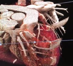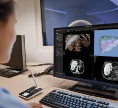
An example of perivascular fat attenuation index (FAI) imaging inside the coronary vascular wall to show areas of inflammation. The technology was created by Oxford University and several experts in cardiac CT imaging say this might be a game changer to improving risk assessments.
There is a promising recent development in cardiovascular computed tomography angiography (CCTA) to analyze plaque to more accurately assess the risk of myocardial infarction (MI), determining which patients to put on aspirin and statins and to have the ability to monitor reversal of coronary inflammation using various drugs. This new plaque assessment technology, the fat attenuation index (FAI), was a big topic of discussion and one of the main take aways from the Society of Cardiovascular Computed Tomography (SCCT) 2020 virtual meting in July.
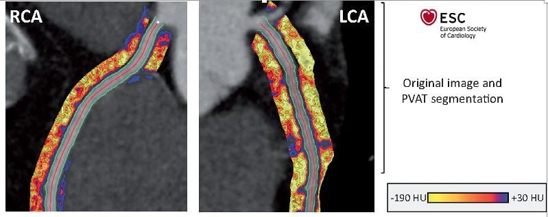
New software developed by Oxford University allows visualization and quantification of the inflammatory response inside the vessel wall, rather than relying on visualization of plaque inside the vessel lumen. The researchers created the perivascular fat attenuation index evaluation as an additional tool to further enhance coronary plaque evaluations and allow more precise classification and prediction, combined with coronary artery calcium (CAC) scoring and intravascular plaque assessment. The FAI color-codes the Hounsfield-based FAI values to make the details more visible and assessment easier.[1,2]
"Over the last four or five years, my group has discovered a new biology," said Charambos Antoniades, M.D., Ph.D., FRCP, FESC, deputy head, Division and Cardiovascular Medicine, director, Oxford academic cardiovascular CT program, consultant cardiologist, Oxford University Hospitals. "When the coronary artery is inflamed, it sends inflammatory signals. These signals begin the process of adipogenesis (the formation of fat cells), lipolysis and edema in the perivascular space of the vessel wall."
To analysis these changes, his team developed the perivascular FAI and automated image analysis software to render images of the vessels from CT DICOM datasets. It uses a radiomics profile that was initially trained to detect tissue inflammation in 1,400 fat biopsies. This is weighted for biological, anatomical and technical characteristics extracted from the DICOM imaging file. It then creates attenuation maps in the perivascular space around the coronary arteries.
"This enables us to understand the degree of inflammation in the underlying coronary artery in the presence or absence of an athrosclortic plaque," Antoniades explained.
CT plaque analysis software has existed for years and can show the various Hounsfield units attenuations to determine morphology of a plaque to show if it is calcified and stable, or soft, lipid-rich plaques, also called low-attenuation plaque (LAP). LAP is known to be unstable and prone to rupture inside the coronaries, leading to thrombus formation that causes heart attacks. However, before the FAI detection system was devised, cardiologists could only guess at the level of vessel inflammation. Antoniades said this new tool will significantly alter our understanding of vessel inflammation as it related to heart attack prediction and prevention.
"I think this might change the way we view coronary anatomy moving forwards," explained SCCT Vice President Ed Nicol, M.D., MBA, FSCCT, Royal Brompton and Chelsea and Westminster Hospitals, London, when he summarized some of the main take away points from the 2020 SCCT meeting.
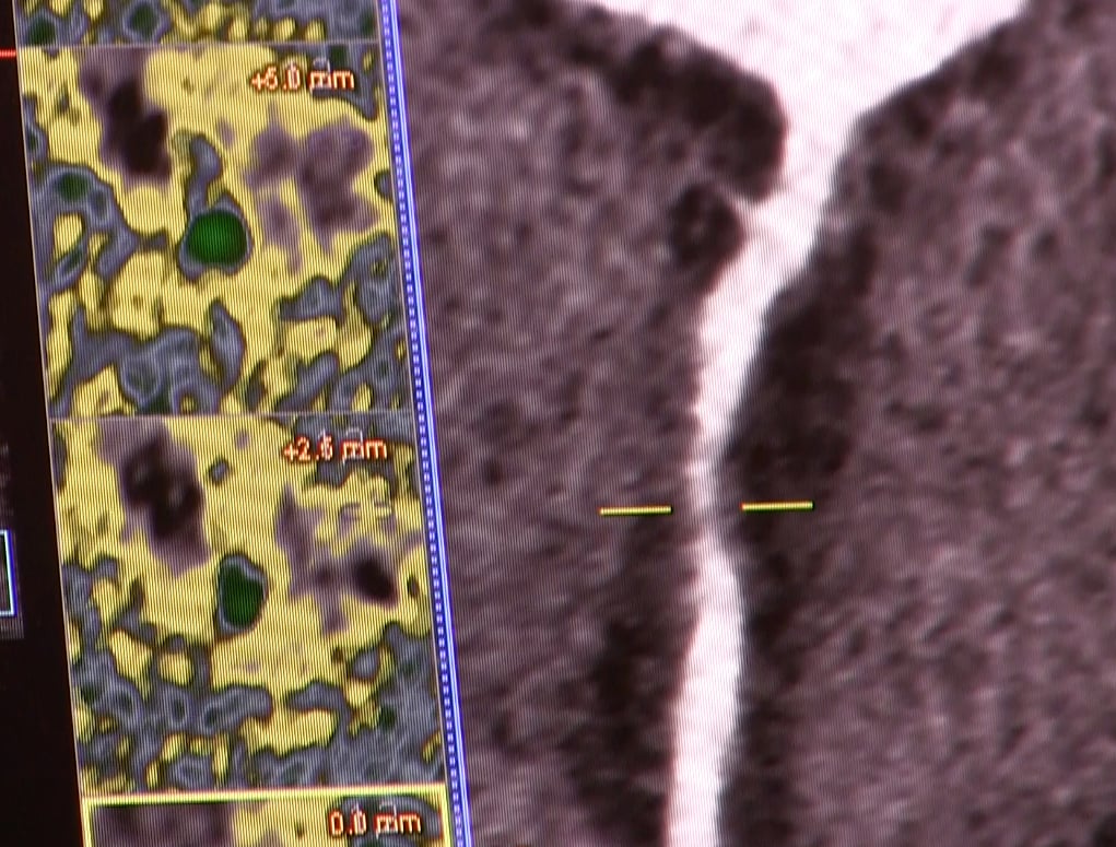
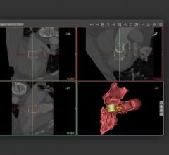
An example of currently available coronary CT angiography plaque assessment software offered by Siemens Healthineers. The right image in a reformatted image to show the entire length of a coronary artery. The yellow marks indicate the location of a plaque stenosis and correlated to the color-coded images on the right, which show and axial view of the artery.
FAI Plaque Assessment Improved MI Detection in Clinical Studies
Antoniades' group led the CRISP-CT study, which tested their FAI technology on about 4,000 patients with up to 10 years followup. It found correlation of 74 cardiac deaths with high rates of FAI.[3]
"It became clear that if your FAI is abnormal, then you have between 5.6 and 9 time high risk of a fatal heart attack over the next 10 years," Antoniades explained.
He said this was more precise than using CT calcium scoring alone and the results showed over and above what you can get from the state of the art core laboratory.
Antoniades said the next step was to add more information on the risk and time line for inflammation, which requires visualization of fibrosis, angiogenesis that includes proliferation of vasa vasorum in the vessel wall, since these are direct indicators vulnerable plaque formation. They did this and combined the data with FAI and found it had an even higher predictive valve. This was tested in the SCOT-HEART Trial: LAP Burden sub-study.[4] This was one of the most talked about trials in sessions at SCCT 2020.
The study used FAI to enhance the assessments and showed low-attenuation, non-calcified plaque and accurately predict future MI. The study found there was a five-fold increase in MI if LAP was above 4 percent.
"It's really the low-attenuation plaque burden that is the strongest predictor MI, above and beyond calcium scoring, and this is really the first time that has been shown. The study had a relatively low number of cardiac events, but a very strong signal in the data," Nicol explained.
Classifying plaque using current technology can be tedious to characterize manually and subject to reader variability, so this study used a semi-automated artificial intelligence (AI)-based FAI plaque quantification software to standardize how the plaque was analyzed. The novel use of AI for automating workflow was also noted by several speakers. The AI-FAI data also helps get more information from CT studies to offer more prognostic value.
"What we see with typical plaque analysis is just the tip of the iceberg. We need new methods to identify the modifiable residual inflammatory risk," Antoniades said. He said it appears FAI offers this additional information.
Nuclear imaging has also been used to image inflammation in the heart. Antoniades explained FAI has a close correlation with nuclear scans to show areas of inflammation.
"What we get from this approach is very similar with what we get from FDG PET," Antoniades explained. In a 2017 study from his group, the uptake of FDG F-18 PET overlays neatly onto of the FAI findings for the same vessels.[2]
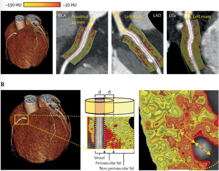
An example of a coronary CT plaque assessment using perivascular fat attenuation (FAI) imaging. It shows a color-coded overlay that indiecates areas of inflammation in the vessel wall, which might be used to improve cardiac risk assessments in the future. Lancet, DOI:https://doi.org/10.1016/S0140-6736(18)31114-0
Next Steps for Using CT Plaque Assessments to Improve Cardiac Risk Stratification
One of the standards of care in heart attack risk prediction is the use of CT coronary artery calcium (CAC) scoring. Many hospitals now offer these low-dose, non-contrast scans as low prices to aid MI prevention screening efforts. Calcified plaques are a sign of previous vessel damage from inflammation or plaque rupture, which is a predictor for future events based on the level of past vessel damage. However, CAC does not take into account the current level of danger from low-attenuation plaques that may rupture, or the level of protection or reversal of plaque formation from a patient going on statins. This is why the findings of the SCOT-HEART sub-study are so important.
Antoniades said another trial, the Oxford Risk Factors And Non-invasive Imaging Study (ORFAN) study, has the objective of recruiting 20,000 patients in phase 1 and 100,000 in phase 2. It will test if the AI-driven FAI assessment can determine outcomes. It also will test how to optimize performance using a deep radiotranscriptomic phenotypic of residential risk with a deep learning algorithm. The goal will be to test personalized risk prediction using CCTA.
Another recent trial, PARADIGM, was asp discussed at SCCT.[5] This study also looked at high-risk plaques in CT scans and found these were the main drivers for subsequent MIs.
Since plaque analysis appears to have a role in improving overall protonic value of CTA, this prompted discussions at SCCT around revising the current CAD-RADS scores. This scoring system is used by cardiologists and radiologists to systematically grade a patient's cardiovascular risk for events based only on anatomical plaque burden severity. It has been suggested that it be revised to be a more detailed multivessel CAD-RADS 2.0 score, which takes into account disease in specific vessel segments and the type of plaques involved to develop more specific risk stratification.[6]
"I think a multivessel CAD-RADS, or a CAD-RADS 2.0, going forwards will be with us in the not to distant future," Nicol predicted.
Better Assessment of Who Should Go on Statins and Tracking Their Impact
FAI assessments can be used in a series of CT scans to track the progress of coronary disease and show the reserve of disease from statins or other drugs, Antoniades said.
He explained the technique was used to evaluate psoriasis treatments in a joint trials between the National Institutes of Health (NIH) and Oxford.[7] The study showed a clear response to drug treatments and a reduction in inflammation compared to the control group.
Several recent studies have revealed aspirin causes bleeding issues in many patients so there is a rethinking of wide-spread prescribing of aspirin for MI prevention. This has generated a lot of interest in identifying the patients who would most benefit and excluding those who are not at high risk for MI. A similar concern remains with statins, where many patients are not sure they want the possible side effects from the drug, so test like this might better define who needs statins and who does not. FAI could also be used to track response to statins.
Related CT Plaque Assessment Content:
Low-attenuation Coronary Plaque Burden May Become Next Big Cardiac Risk Assessment
Top 9 Cardiovascular CT Studies in Past Year
The Latest Advances in Coronary CT Angiography Software
New Cardiovascular CT Technology Entering the Market
Find more content from the 2020 SCCT meeting
Find more content on cardiac CT technology
References:
 May 12, 2020