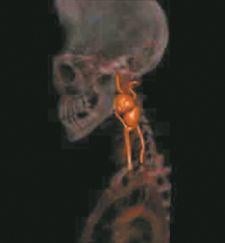
3D image of carotid stenosis.
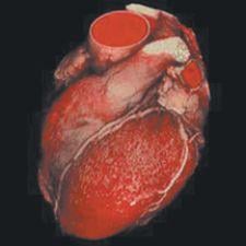
Since the introduction of the 64 multi-slice CT scanner, studies have examined the utility of this technology for diagnosing coronary artery disease (CAD), and cardiologists are rapidly adopting this technology to perform CT Angiography (CTA) for detecting CAD.
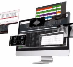
In a clinical setting, however, CTA is only as good as the 3D workstation managing and processing the large volumes of image data CT systems generate. Workstations must have the processing power to move through the cardiac cycle to acquire detailed images, transfer the data and perform 3D reconstructions in real time. Thus, the widespread use of CTA depends to a great extent on the capabilities of 3D workstations.
CTA Adoption on the Rise
As a noninvasive tool for diagnosing CAD and other heart disease, cardiologists are recognizing the benefits of CTA over invasive coronary angiography.
“I can envision CTA as a partial replacement for invasive coronary angiography for the detection of cardiac diseases,” said Michael Ridner, M.D., associate professor, University of Alabama and clinical director, The Heart Center, Huntsville. He notes that the key benefit of CTA is the avoidance of complications due to invasive procedures.
Yet, the value of CTA goes beyond this. “The applications for CTA are very far reaching – it is not just for coronary artery disease but also for valvular heart disease, left ventricular function, cardiac masses and pericardium analyses,” said Ridner. He also sees CTA as a potential replacement for cardiac stress testing and echocardiography. “Many tests, such as stress perfusion imaging, have a lower accuracy rate than CT,” he noted.
3D Workstations Make CTA Possible
Beyond the technical capabilities of multi-slice CT for capturing detailed images of the heart, advances in computer technology and, specifically, 3D post-processing workstations, are the true enabler for CTA studies.
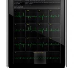
“Without 3D workstations, it would be extremely difficult to perform studies like CTA,” noted Dr. Ridner. He uses TeraRecon’s Aquarius Workstation for reading all 3D CT cardiac, peripheral and cerebral studies, including maximum intensity projections (MIP) and multiplanar reformations (MPR).
At the University of Mississippi Medical Center, David Derr, M.D., associate professor, diagnostic radiology, has a special interest in cardiac imaging studies. His facility uses Voxar Version 6.3 from Barco for volume rendering, vessel analysis and cardiac function. The solution integrates directly into PACS, providing cardiac analysis capabilities on any PACS workstation.
By sending large volumes of data from the CT scanner directly to a Voxar server, “the data loading and processing time is significantly decreased,” Dr. Derr said. “This is particularly important when using functional analysis programs because there are 10 sets of cardiac data that need to be integrated.”
Yasuyuyuki Kobayashi, M.D., radiologist and assistant professor, Department of Radiology at St. Marianna University School of Medicine Hospital in Kawasaki, Kanagawa, Japan, has used Ziosoft’s ZIOSTATION for over five years. Images are transferred from the server to the reading station at 65 slices per second.
Dr. Kobayashi agrees that the value of MDCT increases due to the capabilities of powerful 3D workstations and servers. At St. Marianna, all images are generated with a 0.5mm slice thickness for a daily total of 60,000 to 100,000 slices from two scanners, a 16- and a 64-slice. “By using isotropic data, an accurate diagnosis is possible without the presence of partial volume effect. Plus, the data transfer rate is high and MPRs and 3D reconstructions can be performed in real time, enabling routine use in the clinical setting.”
Most importantly with CT cardiac imaging is the time element, explained Dr. Ridner. Arteries look different throughout the cardiac cycle; therefore, a CTA acquires images of the heart throughout that cycle. “3D workstations must have the processing power to move through the cardiac cycle (for the cardiologist) to analyze the data,” said Ridner. This large volume of data is further increased by the natural motion of the heart. “Viewing this data would be nearly impossible without the additional capability of 4D visualization tools,” Ridner added.
“4D visualization is necessary for the assessment of cardiac function and perfusion studies,” added Dr. Kobayashi. “We must perform both morphological and functional assessment to realize the essence of cardiac diseases.”
Today’s Limiting Factor – CT Scanners
While great strides have been made in CT hardware technology, current limitations still exist. “Currently, the main limiting factor for CT studies is image resolution,” stated Dr. Derr. However, he acknowledges that it is easier to develop new software than to invent new hardware or decrease detector size. “Because coronary vessels are much smaller than other structures we image, in-stent stenoses and/or occlusion remain difficult to evaluate.”
Dr. Ridner agrees that resolution must be improved due to the size of the structures, but he also believes that artifacts in the image, particularly those that are the result of motion or calcifications, are the major stumbling block that clinicians face when interpreting CTA studies. “For more accurate images, CT manufacturers will need to make changes that will increase the speed of acquisition.”
While today’s technology is not sufficient for plaque characterization, specifically determining plaque vulnerability, Dr. Ridner does see advancements in plaque analysis programs. “I do see CTA progressing to help clinicians track therapy over time, to help determine if the therapy is doing what it should,” said Ridner.
Stand-Alone or Integrated
One of the important benefits of a stand-alone 3D workstation is its ability to handle large amounts of data. “That capacity will become more important as data continues to increase logarithmically in size,” said Ridner.
Stand-alone systems have been mainstream for image processing, notes Dr. Kobayashi. Yet, he sees a trend toward networked, or integrated, 3D workstations, primarily to fulfill the need for “image processing methods in a ubiquitous environment at a higher security level.”
While Dr. Derr acknowledges that stand-alone workstations have historically demonstrated better performance because of high-end processor and memory capabilities, “the current generation of hardware significantly levels the playing field for integrated solutions. Rendering can be accomplished just as quickly within the PACS environment as at a stand-alone workstation.”
“The next generation of software will focus on thin-client applications. This will enable access to fully-functional capabilities, even over residential broadband speeds,” said Derr.
The future looks bright for CTA, according to Dr. Derr, who noted, “As vendors continue to push the technology envelope, the diagnostic capabilities of coronary CT will continue to increase.”
 March 06, 2024