Angiography alone for procedural image guidance in the catheterization lab is sometimes not enough, and it is helpful to augment X-ray lumen imaging with intravascular ultrasound (IVUS) or optical coherence tomography (OCT) to visualize the soft tissue morphology.
The Society for Cardiovascular Angiography and Interventions (SCAI) recommends the use of intravascular imaging (IVUS and/or OCT) in addition to fractional flow reserve (FFR) to assess the severity of coronary blockages.[1] It first issued guidance recommendations on the use of these modalities in November 2013. SCAI said these imaging tools can be used to improve patient care, particularly for patients whose heart disease is considered complex or whose disease levels are unclear from angiography.
As use of intravascular imaging increases, there have been a number of technology advances and new vendors entering the market in the past couple years. FFR and intravascular imaging are closely linked for percutaneous coronary intervention (PCI) guidance, so have been combined into one hybrid intravascular imaging system console by St. Jude Medical, Volcano, Boston Scientific and Acist.
Modality Advantages and Disadvantages
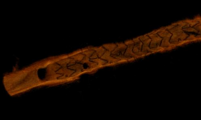
The two imaging technologies are analogous in that they send out energy waves - OCT uses light and IVUS uses sound waves - into the vessel wall and that energy is sent back to the catheter to reconstruct an image. The wavelength of light is much shorter and much faster than sound waves, so OCT has a resolution 10 times greater than IVUS (OCT 10 microns, IVUS 100 microns). In comparison, microscopy has a resolution around 1 micron. St. Jude's newest OCT console also offers the ability to reconstruct 3-D images of the vessel, allowing a new way to visualize vessel segments.
OCT offers clear, photographic quality images, as opposed to grainy, lower-resolution IVUS images that are sometimes difficult to interpret. Fine details such as stent strut apposition and thrombus might be difficult to detect on IVUS, but are very clear on OCT.
However, IVUS has the advantage in its ability to penetrate deeper into tissue to visualize underlying structures. IVUS penetrates 4-8 mm inside the vessel wall, while OCT penetrates about 2-3 mm. If a vessel has significantly remodeled due to plaque burden, the outline of the true lumen disappears on OCT. OCT also requires the use of a saline or contrast flush to clear the column of blood in the imaging area to enable the light from the OCT to image without obstruction.
Volcano has advanced IVUS imaging features to help enhance images and make them easier to interpret. This includes Virtual Histology, which color codes the hardness of materials inside the vessel wall that have a correlation to soft, calcified and necrotic core plaques. Volcano's ChromaFlo imaging shows areas of blood flow in red to help differentiate between static tissue of the vessel wall or stent struts, which can aid in assessing stent apposition, lumen size, dissection and thrombus identification.
New IVUS Advances
Acist Medical Systems gained U.S. Food and Drug Administration (FDA) clearance for its HDi high-definition intravascular ultrasound system (HD-IVUS) in August 2014, becoming the fourth IVUS vendor on the U.S. market. The HDi features an intuitive touch screen with 60 MHz image quality, high-speed pullback, the Kodama HD-IVUS catheter and the Acist HDi console. The vendor said its pullback is 20 times faster than other currently available IVUS systems, lowering the risk of catheter-induced ischemia by reducing pullback time from minutes to seconds. The system is also the first to offer FFR using a rapid-exchange catheter.
In August 2014, Medtronic entered into an agreement with Acist to co-promote the rapid exchange FFR RXi and HDi technologies in the United States.
Volcano launched SyncVision co-registration system for angiography and IVUS at the 2014 Transcatheter Cardiovascular Therapeutics (TCT) meeting. The software features a live, online image processing workstation for coronary catheterizations that allows physicians to simultaneously navigate on both an angiogram and an IVUS image in a single correlated view. This allows pinpointing an image of a vessel cross-section showing a specific lesion and matching the vessel location on the angiographic image.
Boston Scientific launched its new Polaris IVUS imaging system that is FFR-capable in July 2014. It also announced a partnership a month later with Asahi Intecc to develop a new, differentiated FFR wire. The joint project focuses on creating a device intended to improve handling compared to existing FFR wires. Boston Scientific anticipated commercializing its FFR wire in 2015.
The Polaris system is designed to be smart, fast and accurate, with a workflow and interface designed for greater ease of use than previous Boston IVUS systems. It uses a modular design and supports the planned release of a new FFR wire, a new family of IVUS catheters, enhanced software features and better system control tools.
Boston Scientific launched its next-generation OptiCross IVUS catheter in July 2013. It was designed to offer better deliverability and higher resolution imaging than previous Boston Scientific IVUS catheters. It has a low-profile delivery system with a 5 French guide catheter compatibility; a shorter, tapered tip; a bi-segmented catheter shaft; and a redesigned catheter hub for ease of connection.
Near-infrared Spectroscopy to Identify Vulnerable Plaques
The Infraredx TVC Imaging System combines IVUS with near-infrared spectroscopy (NIRS) to analyze the chemical makeup of plaques. The system shows a halo ring of color around the IVUS image, with yellow indicating plaque and red representing lipid core plaques. The TVC Imaging System is U.S. Food and Drug Administration (FDA)-approved to identify lipid-core plaques that may cause heart attacks. Identification of such plaques would be a major step toward the development of percutaneous coronary intervention (PCI) as a means to prevent coronary events. The company is in the process of further validating the technology's ability to detect vulnerable plaque with a series of trials.
In April, Infraredx announced the enrollment of 1,000 patients in the Lipid-Rich Plaque (LRP) Study. It is a prospective, multi-center clinical trial designed to identify a correlation between lipid-rich plaques detected by the TVC Imaging System and the occurrence of a cardiac event within two years.
In December 2014, the results of the THEROREMO-NIRS Study were published in the Journal of the American College of Cardiology (JACC), showing NIRS can correctly identify lipid core-containing plaques. In the study, patients who presented with symptoms associated with limited blood supply to the heart also underwent NIRS imaging to evaluate the lipid core burden index (LCBI) in an artery that was not directly implicated in causing their symptoms. The results demonstrated that patients with an LCBI ≥ 43 in a non-culprit artery had a fourfold risk of major adverse cardiac and cerebrovascular events (MACCE), such as heart attack or stroke within the following year. In addition, the study concluded that non-culprit vessel LCBI reflects vascular vulnerability of the larger coronary tree.
The study, a sub-study of the European Collaborative Project on Inflammation and Vascular Wall Remodeling in Atherosclerosis (ATHEROREMO), was a prospective, single-center, observational study that enrolled 203 patients referred for coronary angiography due to stable angina or an acute coronary syndrome (ACS), a combination of symptoms resulting from the blockage of blood supply to the heart. NIRS imaging was performed and an LCBI measurement was obtained for a pre-defined segment of a non-culprit coronary artery that was at least 40 mm in length and with < 50 percent stenosis confirmed by angiography. The primary endpoint was the incidence of MACCE, defined as all-cause mortality, non-fatal ACS, stroke and unplanned coronary revascularization during one-year follow-up.
Data from the ongoing Lipid-Rich Plaque Study and PROSPECT II/ABSORB Study will also provide further data on the use of NIRS.
OCT Advances
St. Jude Medical gained FDA clearance in October 2013 for its Ilumien Optis PCI optimization system. This is the newest generation of the company's combined OCT intravascular imaging and FFR system. It offers unique software that enables real-time 3-D image reconstructions of vessels from OCT datasets, making it easier for physicians to visualize the area they are treating. The technology is similar to the use of computed tomography (CT) or magnetic resonance imaging (MRI) 3-D image reconstructions. The system also has automated measurements and stent planning software tools.
The Ilumien Optis offers twice the OCT resolution of the earlier generation. The system uses the Dragonfly Duo imaging catheter, which offers faster, longer pullbacks to assess artery segments in less time.
Avinger became the second interventional OCT device vendor in the United States with the November 2012 launch of its Ocelot OCT-enabled chronic total occlusion (CTO) crossing catheter.
Avinger received European CE mark approval in September 2013 for the Pantheris directional atherectomy catheter that is combined with real-time OCT intravascular visualization to treat peripheral artery disease (PAD). In July 2014, Avinger said the first patients were enrolled in the VISION trial, a global investigational device exemption (IDE) clinical trial approved by the FDA to evaluate the Pantheris catheter.
1. Amir Lotfi, Allen Jeremias, William F. Fearon, et al. "Expert Consensus Statement on the Use of Fractional Flow Reserve, Intravascular Ultrasound, and Optical Coherence Tomography." Catheterization and Cardiovascular Interventions 00:00–00 (2013).

 November 14, 2025


