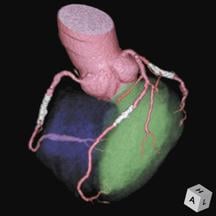
Ziosofts multi-volume fusion capability enables clinicians to show spatial relationships between various anatomical objects.
Volumetric data achieved from multislice CT and MR systems provide valuable insight for clinicians, especially within the cardiac arena where the number of images required to diagnose and treat a single patient can range from tens to thousands. All this data, however, comes with a caveat: How to process it into manageable, interactive information that fully captures the imaging modalities’ potential.
Stand-alone workstations used to be a clinician’s only choice for rendering imaging data into 3D, thereby reducing the number of images needed for review and increasing interpretation accuracy. Today, advanced visualization software applications have the ability to process and display large image data sets, and at the same time allow clinicians to manipulate CT and MR slices into 3D and 4D images to create detailed models of anatomy. The technology can be used to plan treatment options, routes through vessels and to determine the size and types of devices needed for a procedure.
“Cardiac imaging is very complex, particularly the visualization of the human coronary arteries,” said Michael Poon, M.D., director of advanced cardiac imaging, department of radiology, Stony Brook University Medical Center, Stony Brook, NY.
“The 3D advanced visualization tool takes hundreds of 2D images and reconstructs them in a 3D fashion, allowing you to look at the entire structure — the heart —- in one view. It also allows you to manipulate the image for infinite viewing angles, change the contrast and change the image orientation,” he explained.
Thin is in
One of the biggest downfalls of a stand-alone 3D workstation — or thick-client — model, according to Terry Chang, director of marketing for Ziosoft, is that raw images have to be “pushed around the network.” With true thin-client architecture the processing horsepower is centrally located and results are streamed to the client workstation.
“Hospital networks are getting faster but they can only handle so much traffic and if you have to push these images around the hospital, it is very inefficient, it bogs down the network and it discourages sharing of that information among different users,” explained Chang. “Now that 3D’s reach extends not only to radiologists, but also cardiologists, surgeons, oncologists, physicians in the ER and referring physicians, it truly is an enterprise-wide tool that needs to be everywhere. Physicians want access to a patient’s 3D image and they want to not only view it on PACS, but they want to interact with it,” he added.
Advanced visualization tools provide physicians with this interactive review of images, allowing them to manipulate the data for quicker, more accurate diagnosis and treatment planning. And thanks to thin-client architecture, physicians have immediate access to these capabilities anywhere within the enterprise.
Market offerings
Ziosoft’s Ziostation provides a full suite of advanced 3D analysis tools and features a customizable user interface and action macros to maximize efficiency across various user types. The company boasts a true thin-client solution that supports both robust and less robust applications including: 3D, 4D, MIP, MPR, CPR, multimodality viewer, multivolume fusion, vessel analysis, coronary analysis, colon analysis, PET/CT viewer, CT cardiac function analysis,* calcium scoring* and reporting functions. (*Currently not cleared for sale in the U.S).
Its recently FDA-cleared CT brain perfusion application provides brain perfusion functionality to clinicians as a thin-client application using standard commercial hardware. It aids in stroke assessment by providing a color map of cerebral blood flow and other perfusion-related parameters from CT images of the brain. Image manipulation tools as well as measurement tools such as cerebral blood volume, blood flow and mean transit time help physicians — whether in the hospital or at a remote location — assess the type and extent of a patient’s cerebral perfusion disturbance.
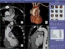
TeraRecon offers what they refer to as a paradigm shift in 3D visualization with the Aquarius iNtuition Client that delivers workstation-class applications to regular PCs or PACS stations throughout the department and enterprise through the convenience of a thin-client platform. Aquarius iNtuition boasts one seamless solution for advanced image management needs by delivering the specific high-end tools and defined workflow steps anywhere they’re need within the enterprise.
“The most common clinical application of advanced visualization today is evaluation of coronary artery disease,” said Dr. Poon who works with both the Aquarius workstation and Aquarius iNtuition thin client. “It is extremely important to be able to interact and manipulate with the image data because the human coronary is somewhat hidden under structures. That is what is so valuable about the Aquarius — it allows you do virtual dissection, you can remove or put back structures. It also has the capability of memorizing case-specific workflow, automatically going through the steps you normally perform on a routine case.”
Dr. Poon also appreciates the Aquarius’ ease of use and intuitive platform, features that led him to choose it as a teaching tool at the university. And with the number of volumetric imaging cases growing, the need for this type of advanced diagnostic image manipulation becomes even more important, he says.
Among the host of available robust Aquarius tools Dr. Poon regularly uses are the TVA (time volume analysis) tool to calculate ejection fraction and the easy-to-use calcium scoring tool. A very powerful application used for more complicated cases is 4D data review, which because of the amount of motion-related artifact associated with cardiac CT, allows you take look at an image over time, he says.
Yet, as pleased as he is with today’s advanced visualization capabilities, Dr. Poon does see the need for further evolution. He would like development in the area of data transfer as well as artificial intelligence that would allow users to select out the images that are more important. He also sees the need for a strategy that provides the physician a simpler way of dealing with large datasets.
Flux within the advanced visualization market is likely to due to evolution and demand. Current participants, aside from TeraRecon and Ziosoft, include Vital Images, Visage Imaging, Merge Healthcare, FiatLux Imaging, Calgary Scientific and more. In November 2008, Barco sold off its advanced visualization business and solutions, known as Voxar 3D and Voxar 3D Enterprise, to Toshiba’s newly formed subsidiary, Toshiba Medical Visualization Systems (TMVS).
Balancing act
For physicians on the go, Ziosoft’s Web client application, Ziostation Web, provides 3D analysis and real-time collaboration tools on a secure Web site without the need for any proprietary or specialized hardware. It expands the reach of the Ziostation thin-client 3D system to virtually any computer with a Web browser, giving authorized users, no matter where they are, access to original and saved images and reports.
Similarly, TeraRecon’s AquariusWEB Client delivers volumetric image review anywhere, and is designed to run natively and securely within a Web browser with no software download required. An automatically generated email with a URL link can be sent from one person to another, and the study of interest can be opened by the recipient by simply clicking the link.
According to Chang, the Web client option is growing in popularity because more and more physicians want the accessibility to images wherever they are in the world. While they are willing to sacrifice functionality for accessibility, most draw the line at compromising image integrity. As a result, choosing between advanced visualization market offerings becomes a balancing act. Based on their specific needs, facilities must find the solution that provides the best balance among functionality, bandwidth (accessibility) and image integrity.
 May 12, 2020


