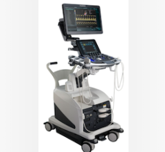
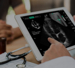
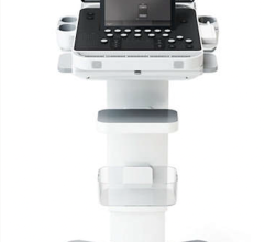
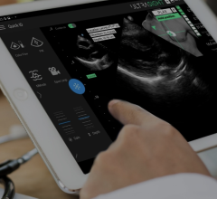
This comparison chart covers dedicated cardiac ultrasound scanners specifically intended for performing cardiac or vascular imaging studies. General purpose systems with extensive cardiac scanning options are also included.
Cardiac ultrasound scanners are ultrasound scanning and image processing systems designed specifically for real-time, noninvasive imaging of heart structures. They are used to detect such conditions as mitral and aortic stenosis and insufficiency to determine the extent of damage from suspected myocardial infarction and to diagnose congenital cardiac defects — such as patent ductus arteriosus and transposition of the great arteries. Cardiac ultrasound can also be used instead of cardiac catheterization to monitor ventricular function. Transesophageal echocardiography (TEE) is most commonly used in surgery for detecting myocardial ischemia and monitoring cardiac output. This intraoperative use of TEE allows analysis of regional cardiac wall motion, in which abnormalities have been shown to develop within 15 seconds of coronary occlusion.
Vascular ultrasonic scanning gives the physician profiles of arteries and veins throughout the body. It is used to diagnose atherosclerotic obstructions, occlusions, disease and incompetence by means of a 2D, real-time image of the organ or vessel, as well as a profile of blood-flow velocity through the area being examined. In many cases, vascular ultrasonic scanning systems prevent the need for contrast arteriography, which requires vessel cannulation, contrast media injection and ionizing radiation exposure. Vascular ultrasound imaging is the primary screening method for deep vein thrombosis (DVT). Many ultrasonic scanning systems that are marketed primarily for cardiac and vascular applications can be used for other applications; however, additional transducers or software may be needed.
Various probes of different ultrasonic frequencies are available. For diagnostic imaging, frequencies from 2 to 30 MHz are typically used, while frequencies of 5 to 15 MHz are considered optimal for vascular scanning. Probes that generate higher frequencies produce shorter wavelengths and narrower beams, which improve resolution; however, higher-frequency sound energy is more readily absorbed by tissue and the usable depth of penetration is decreased. Many systems now have broadband probes, which have larger frequency ranges than traditional probes and offer combinations of deeper penetration and higher resolution.
Various modes are available for displaying the returning echoes. B-mode (brightness-modulated mode) is the scanning system’s basic imaging mode. B-mode produces a real-time, 2D image that represents a cross-sectional slice of the area under study. M-mode (motion-mode) uses a fixed position pulsed beam to produce a moving display of a single scan line over an interval of time. Used almost exclusively in cardiac studies, M-mode produces a graphic display of a moving structure (e.g., the cardiac valve over several heart beats.) Simultaneous display of M- and B-modes is particularly useful when examining dynamic structures such as the heart.
Cardiac ultrasound scanners use Doppler to determine the direction and speed of blood flow. Most scanners include spectral Doppler, either continuous wave (CW) or pulsed wave (PW). Spectral Doppler includes a spectrum analyzer to display frequency shifts plotted against time with grayscale intensity varying with the received signals’ strength or amplitude. Color Doppler imaging of myocardial tissue to show motion and assess myocardial viability is being researched for applications in stress echocardiography evaluations of heart abnormalities (e.g., Wolff-Parkinson-White-Syndrome) and reperfusion therapy.
Some manufacturers offer 3D ultrasound, which involves volume-per-second acquisition and display for volume measurements, improved imaging presentation and volume-of-interest studies. The 3D images can be produced by direct online 3D acquisition with a transducer scanning a volume instead of a slice of the tissue. An advantage of 3D ultrasound is that it can simulate intraoperative visualization.
Expanding vascular applications include guided sclerotherapy; assessment of pelvic venous congestion, sapheous insufficiency, saphenofemoral reflux and perforator disease and imaging of the lower-extremity DVT.

 October 09, 2025


