Mannudeep Kalra, M.D., radiologist, Massachusetts General Hospital
Foremost on the minds of many radiologists today is how to reduce radiation dose when using computed tomography (CT) without compromising image quality.
Imaging Technology News took the questions to two experts in the field, Keith Dreyer, D.O., Ph.D., vice chairman of radiology, Massachusetts General Hospital, and assistant professor of radiology, Harvard Medical School, and Mannudeep Kalra, M.D., radiologist, Massachusetts General Hospital.
Dreyer emphasized the use of standards-based practices with the help of decision support technology for reducing patient exposure to radiation dose, and Dr. Kalra recommended several techniques for lowering CT dose while actively scanning.
Protocols, Decision Support Help Reduce Dose
“There are three immediate ways to reduce the radiation burden of computed tomography (CT),” Dreyer said.
“First, review your CT protocols such that the radiation dose per examination is optimized, and minimized where appropriate. Oftentimes institutions do not review their imaging protocols with the implementation of new equipment and as such are more likely to deliver a higher radiation dose than what is necessary for optimal image quality.”
“Second, ensure proper utilization of computed tomography,” Dreyer said. “The inappropriate ordering of imaging procedures can occur from the variability between referring physicians — some order too often, some too infrequently. By using standards-based ordering decision support, referring clinicians can enter a patient’s symptoms and qualify the effectiveness of their exam choice through real-time appropriate medical image ordering guidance. For example, Nuance’s Radport is a decision support system that guides appropriate diagnostic image ordering to reduce ordering time by up to 80 percent, manage high-cost utilization and improve patient care through appropriate imaging. The result is that over-utilization due to physician variability can be considerably reduced.”
There are always alternative to CT, Dreyer notes. “Third, order alternative (nonionizing radiation based) modalities such as MRI and ultrasound. Again, with the use of a decision support system, alternative modalities can be identified, over-utilization avoided and orders rerouted to more appropriate procedures for patients,” Dreyer said.
Adapt to Clinical Indications
Protocol rules out the odds of unnecessarily exposing patients to excess radiation, but it is only the first step. Kalra believes a couple of key steps include following clinical indications, then to adapt the scanning protocol to the clinical indication and to adapt the scanning protocol to the size of the patient.
“The first thing is indication. If a scan is indicated, then the dose is justified to obtain required information,” said Kalra “The dose question first comes up with the appropriateness. The American College of Radiology (ACR) laid down their appropriate criteria, which Dr. Keith Dreyer used to make decision support trees to help physicians decide what test, CT or not, is good for the patient.”
Kalra points to a recent article published in the New England Journal of Medicine, “Computed Tomography — An Increasing Source of Radiation Exposure” by David J. Brenner, Ph.D., D.Sc., and Eric J. Hall, D.Phil., D.Sc. Brenner and Hall suggest that about one-third of the scans performed in the United States are being done without good clinical indications, and information that is expected from these scans can be obtained by other tests that do not expose patients to radiation or only use very minimal radiation, for example, radiography and ultrasound.
“Once it is determined that there is a valid indication for CT scanning, the indication for CT should help the radiologist to determine appropriate CT protocol which governs the associated radiation dose to some extent,” said Kalra. “So, the first step on dose reduction starts with the radiology department educating their physicians about appropriateness of the exam.
After the indication, the second most important step towards managing radiation dose is recognition of the fact that children are more vulnerable to the risk of radiation induced cancer, Kalra said. “Roughly speaking, young children are about seven times more at risk of developing cancer from the same radiation dose as an elderly person. Girls are at much higher at risk than boys at the same age for the same dose. Thus, it is essential to ensure that CT protocols and techniques give lower radiation dose to a younger person, particularly children, compared to an older individual.”
Kalra expounds upon other notable studies recommending techniques and steps to take in order to reduce radiation dose in children.
“The first principle of dose reduction in a radiology department is that children should always get lower dose than adults for the same protocol in the same body region. But, for instance, if you’re doing head CT in an adult or chest CT in a child, your head CT in an adult is allowed to have lower dose than a chest CT in a child,” Kalra said.
There is a latent period following radiation exposure and the time it takes for the radiation effects to develop. The latent period for development of cancer following low level radiation dose is variable and can be as long as 10-30 years, Kalra said. “For a person getting a CT scan at age 60, he is unlikely to develop cancer in his remaining lifetime, but children have much longer to live,” Kalra indicated. "Children’s cells are not only more susceptible to radiation but they are also likely to live longer than adults, reinforcing the point that each department should first focus on lowering dose with children. Children should always get lower dose than adults for the same protocol in the same body region.”
Kalra offered the following techniques for reducing cardiac CT dose:
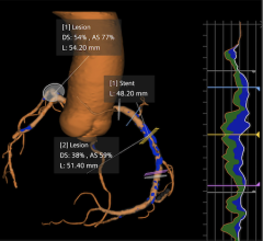
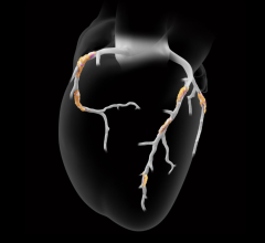
Cardiac CTA
Although we only scan 15-20 cm of the chest for most cardiac CT exams, particularly coronary CTA, the doses are relatively higher because we’re trying to the look at very fine structures with overlapping data acquisition. And because you are acquiring and matching the data with the cardiac cycle or the motion of the heart, you are overlapping the data. That is why you have higher dose with cardiac scanning because you need higher image quality. You need higher resolution and thinner slices, and those are things that contribute to higher dose for cardiac.
ECG Pulsing
One of the ways of reducing dose for cardiac CT is called electrocardiogram (ECG) pulsing (or ECG controlled tube current modulation). If you are looking for coronary arteries and the heart is constantly moving, what we do is segment the heart in order to obtain a measure of the coronary arteries when they are least moving. We only want to acquire coronary arteries when the heart is relatively resting, which is the diastolic phase.
We hook up the tube current with the ECG signal that we obtain when we are scanning the patient. We increase the tube when we are over that resting phase or the diastolic phase and then we drop it down during the systolic phase, or when the heart is moving very rapidly. So, we pulse the tube current based on the ECG signal.
If the systolic phase is longer than the diastolic phase, this occurs when the patient’s heart rate is too high, this results in little dose reduction. But on the other hand, if you have a patient with 60-65 beats per minute heart rate, you can actually obtain 30-50 percent dose reduction using this technique. So, the amount of dose that can be reduced using ECG pulsing depends on the patients’ heart rate.
Prospective ECG Triggering
More dose reduction can be obtained through prospective ECG triggering. What is the difference between prospective ECG triggering and the ECG tube current modulation? In prospective ECG triggering instead of dropping the tube current during the phase when the heart is rapidly moving, we turn off the tube current to obtain much higher dose reduction. This technique is commonly employed when we are doing coronary calcium scoring and to do coronary CT angiogram also, if the patient’s heart rate is stable and slow. Because one of the pitfalls that can occur is the patient suddenly has a premature beat or has an arrythmias. Then you don’t want the tube to be off when the heart is relaxing. So that is the pitfall. You need to have a regular and slow heart rate. You need a regular and slow baseline heart for this to be used and you can up 80 percent dose reduction using prospective ECG triggering. So perhaps the lowest dose can be performed using ECG pulsing, as low as 1-2 milliSieverts of dose for coronary calcium scoring and coronary CT angiogram, using this technique.
Automatic Pitch Adaptation
The third technique for cardiac is the automatic pitch adaptation. Most scanners have this. When the heart is moving fast, they make the scanner go faster, and there is less overlap and higher pitch during scanning. When you do that, you reduce the dose.
With the dual source multidetector CT (MDCT) the extent to which you can change the pitch is much greater. With a single source 64-slice scanner you can only change the source from 0.2–0.35. But with dual source you change the pitch from 0.2 to 0.55 – almost double. As a result, as the patient’s heart rate goes higher the pitch can go higher. As the pitch goes higher you get less overlaps, therefore, your dose is getting lower.
If you look at the ECG tube current modulation, it had the opposite effect. When you have a higher heart rate your dose reduction with ECG pulsing was lower.
With a faster heart rate, you have higher pitch and therefore lower dose. That means for a dual source scanner, perhaps you want patients who have a higher heart rate, so you don’t have to give beta blockers to the patients.
With dual source you don’t need the beta blockers because the scanner is faster, and you can cut down the dose using the pitch adaptation.
Lower Kilovoltage
The last thing about cardiac CTA is using lower kilovoltage (kV). If you use 100 kV instead of the 120 kv that is conventionally used, you can drop the dose by 30–35 percent. Reducing the dose by reducing the kV, you increase the contrast in the image and therefore you can tolerate higher volumes when you have higher contrasts. That’s one of the rationales for going to a lower kV versus a higher kV.
This comes with a huge caveat. If your patient’s size is above average, the image quality can suffer. So then if the user wants to use lower kV, he has to look at the patient’s size. For example, with a 150-pound woman, although she is a smaller than average patient, if she has large breasts, then she is a large patient for CT for the heart. So, the regional size of the body or large region of interest is very important and avoid doing lower kV with large size patients or patients with a large region of interest.
Whatever scanner you have, you need to follow indications first, second adapt the scanning protocol to the clinical indication and the size of the patient.
What should be basic image quality has to come through Radiologists efforts. Vendors don’t know about the clinical indications, and the radiologists are not as up to date on the technological capabilities. There is awareness that radiation is a concern, but this requires education amongst radiologists.
Automatic Exposure Control Technique
One of the most important methods that has been developed is the automatic exposure control technique. This technique, if used appropriately, will allow the radiologist or cardiologist to reduce does in children by 30-50 percent. The clinician selects an appropriate level of image quality. The system then calculates the size of the child or the adult — it can be used in adults also — and automatically uses the lowest possible dose obtain the desired image quality. It adapts the tube current to the patient’s size based on what image quality the clinician has specified.
The Color-Coded Technique
Donald P. Frush, M.D., professor of radiology and pediatrics, director, division of pediatric radiology, a leading pediatric radiation dose expert from Duke University School of Medicine, has reported another innovative method for reducing dose to children, a color-coded technique for adapting the dose to the child’s size. GE Healthcare scanners have this technique on their console. That technique uses the patient’s weight and a certain kilovoltage and tube current to obtain certain dose. If the patient is very small, it is going to use lower tube potential and current to obtain lower dose studies. If the patient is heavy or older, it uses a relatively higher tube current and higher tube potential, while still maintaining doses below the adult doses.
Inspired by Frush’s seminal paper on color coded pediatric protocols for dose optimization, at the 2007 RSNA we presented a paper in which he went one step further than Frash. We said ok, Frash’s protocol only relies on patient size to reduce the dose. He said the dose should not only be tailored according to patient size, but also whether the patient had a CT scan for the same indication.
For example, a five-year old has a CT of the chest to look for pneumonia. And they find the pneumonia, and they say come back in six to eight weeks to see if it has gone or not because the treatment is going to differ if it has gone. If it’s gone, then you don’t need to do anything. If it is not gone, then the doctor might put a scope in to see if there is a foreign body. Often you get a follow-up CT. Or if the child has a cancer, like lymphoma, he will have a baseline CT. After six or eight weeks, the patient will have a follow-up scan.
So, apart from just relying on the size of the child to predict the dose, we should also rely on two other things: one is whether the patient had a prior CT. The doses of CT are accumulative. The radiation that you accumulate over your life is important. The first 100 or so units of radiation are added to the new 100 units of radiation, and you get a cumulative effect. Therefore, we said, not only do you need to see the size of the child, but also whether he has had CT scans before for the same indication. For example, if a child had a CT scan for pneumonia a month ago, you don’t need to give him the same dose just to see if it is gone or not. You need to cut down the dose.
Dose should be adapted to the size of the patient, the number of prior CT studies and to the clinical indications. I think these principles should also be applied to adults.
Automated Exposure Control
Then we use automated exposure control to adapt the tube current to the patient’s size and we also change the kilovoltage of the tube potential. With all of these factors, in order, we optimize the dose for the patients.
Now, with the adults, you can also use the automated exposure control technique because they come in different shapes and sizes. These techniques adapt the dose to their size and shape. One important part of automated exposure control is how you select what image quality should be obtained from the technique. It is like the Achilles heel in CT – what is the optimal imaging quality you can get because the optimum quality not only depends on the patient’s size, but also depends on the indication of the study.
For example, if there is a lesion in the liver, you require a relatively better quality image. If the lesion is a stone in the kidney, you can accept a lower quality or a noisy image. This is something that the radiologists need to decide based on the clinical indications and level of comfort to assess low radiation dose images. The automated exposure control is only automatic to a certain extent. It will only adapt to the patient size. It won’t look at the clinical indication unless the radiologist sets the level of image quality. Inappropriate selection of superior image quality can actually increase the dose with the automated exposure control technique. That is why automated exposure control technique is only automatic to a certain extent to make sure that proper levels are selected.
Lower Tube Current
There are other techniques for lowering dose in children and adults. These include using a lower tube current, and that would be applicable to any protocol. For example, using a lower tube current, using automated exposure control, using a lower tube potential or kvp, using a higher pitch or using a better detector profile or geometry.
For example, a 4-slice scanner has higher dose than 64-slice scanners. The efficiency of a 4-slice scanner is around 70-80 percent. The efficiency of a 64-slice scanner is around 90-95 percent. So detector geometry also affects dose. And then reducing scan lent or body regions that are scanned helps you reduce dose.
Fewer CT Phases
It is also important to center the patient right in the center of the gantry. If you don’t center the patient right in the center of the gantry, you can increase the dose by 11-15 percent. This is a very common error.
The other thing is how many times you scan the same body part. By reducing the number of passes that you take of the same part of the body, you will reduce the dose. Reducing the number of phases in CT can help you reduce the dose.
Noise Reduction Filters
There are also the noise reduction filters. Noise reduction filters is another upcoming technique, which has been around for a while. Several vendors have them on their systems and others sell them as third-party vendors. In this technique, you take the image that has been acquired at a lower radiation dose and they electronically manipulate the image to match the image that is required at the usual higher dose. They are post processing image-based software. Now we do need more evidence to see whether they are associated with any artifacts, but this is a potentially promising way of looking at dose.
There are other studies where you can use these noise reduction filters to reduce the dose by as much as 50 percent.
These are the general parameters you can use to reduce dose. Several studies show the noise is lower, but that the image is sufficient to make a diagnosis. As long is the image noise is acceptable, radiologists or cardiologists should use less radiation.
Reimbursement for CTA
The reimbursement cuts will affect the smaller institutions or where people can’t pay out of pocket will see a decrease on the use of CT for coronary CTA. Most places in the United States are not reimbursing for CTA.
I don’t think the slice war, as they call it, is inappropriate. The push by the vendors is in the right direction because CMS says there is not enough evidence [to support reimbursement for CTA].
There are several limitations in cardiac imaging. Spatial resolution and temporal resolution is much lower than invasive catheter-based coronary angiography. Perhaps with scanners that are much faster, there will be quick outbursts of evidence to do away with invasive coronary angiogram. If [manufacturers] are able to improve the temporal resolution, I think coronary angiogram would emerge ultimately with better evidence needed by the third party payers to reimburse coronary CTA studies.


 February 02, 2026

