The SOMATOM Sensation 64 from Siemens is designed to deliver 64-slice sub-millimeter
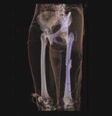
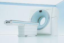
Today’s patients present to the emergency department (ED) with a slew of complaints and injuries ranging from a chronic cough to a sudden life-threatening head trauma. As such, the ED is no longer reserved for those patients with dire life or death circumstances, as the standards of emergency care apply to all patients and handle a larger caseload. With this changing dynamic, physicians rely heavily on imaging modalities to quickly triage patients and uphold emergency standards of care in providing an accurate, complete and immediate diagnoses. In many respects, the ED is becoming more of an emergency radiology department or ERD. Radiology takes over the ED A leader in the ED to ERD trend is Brigham and Women's Hospital, an affiliate of Harvard Medical School. Stephen Ledbetter, M.D., chief of the Emergency Radiology Division at Brigham and Women’s, explains that a dramatically increased use of the emergency department by patients has led to the creation of their emergency radiology division. “That trend is going to continue because of demographics - which includes an aging, more diverse and uninsured population,” he explained. “Emergency medicine has become a full-blown specialty just like neurology or plastic surgery, and like other specialties it now needs services that are coordinated and aligned with its clinical service. Emergency medicine needs a dedicated emergency radiology section to ensure the smooth delivery of care. Emergency radiology is a different vehicle for the delivery of emergency medicine services. It is no longer a delivery vehicle that tries to obtain services from five, six, seven or eight subspecialties of radiology. It needs those subspecialty services coordinated under one umbrella.” Specifically, an ED develops into an ERD once it is delivering emergency radiology coverage 24/7 and has around the clock access to imaging tools to support it. “Historically, emergency radiology was on-call radiology and that’s why, in the past, there was no emergency radiology department,” Dr. Ledbetter said. “The old model included extended hours, multiple consecutive hours and working alone with limited resources. There was not a dedicated section to provide and foster oversight, advocacy and planning. Emergency radiology takes that historic model and flips it on its head; it has moved from an on-call model to a service model.” The ED provides around the clock radiology coverage by having constant access to the latest technological advancements in imaging. Although one may have equal access to computed and digital radiography (CR/DR), three-dimensional computed tomography (CT) and magnetic resonance (MR) imaging , several factors come into play when determining which modality is best suited for a given patient. While CT and MR provide the best imaging results, CR and DR are the most frequently used in the ED, because they are fast, less expensive and expose patients to the least amount of radiation. Accuracy, specificity drive CT and MR in the ED With the heavy reliance on CR/DR for many indications presented in the ED and with its convenience, quick assessment and low cost, what then prompts physicians to order CT or MR? According to C.E. Ballard, M.D., emergency department staff physician at Baptist Medical Center in Little Rock, AR, “The chief complaint for a plain CR/DR image is an indication of an acute, chronic or exacerbation of a chronic illness, a chest X-ray or a long bone X-ray. If we get that X-ray and it’s unclear, then frequently the radiologist may indicate a CT or MR.” William R. Reinus, M.D., MBA, FACR, vice chairman of the Department of Radiology at Temple University Hospital concurred. “If we can't figure out what’s wrong, it's better to spend money upfront than have something disastrous occur,” he said. “With the legal repercussions that exist, most people are inclined to be fairly liberal in the number of studies we order.” Because of the advancements in technology, specifically in CT, Dr. Reinus uses Siemen's SOMATOM Sensation, a 40- and 64- slice CT using the latest z-Sharp technology, which provides the highest routine isotropic CT resolution of 0.33 mm voxel size and complete elimination of spiral artifacts. Siemen's SOMATOM Sensation is able to scan a thorax in four seconds and freeze the heart's motion using the industry's fastest gantry rotation time of 0.33 seconds. According to Dr. Reinus, “The goal that we need to work toward is to get the maximum amount of imaging with the least radiation exposure. To that end, it’s certainly been the progression of CT to do more, faster, but at some point people are going to have to think about doing survey-projectional images and cross-sectional images. The patient will need be in the scanner and need a topogram in different planes in which the patient only goes through the scanner once with contrast and once without.” CT has become quicker and more accurate, however, each new advancement and improved efficiency prompts one to dream even further into the future of imaging. Another imaging tool on the cusp of more widespread use is coronary CT angiography (CCTA). It can rapidly and accurately assess chest pain by providing an inclusive set of images for evaluating the three main causes of chest pain: a pulmonary embolism, aortic dissection and coronary artery stenosis. The CCTA, using the latest 64-slice multidetector computed tomography (MDCT) systems, enables visualizing the entire chest of all three areas of suspicion. This type of assessment can be performed in about 12 seconds; however, the 256-slice system now being tested can scan the heart in 1.5 seconds. MR sets gold standard, yet not fast enough Each physician said even though MR has exceptional image quality, it is the least utilized in emergency situations. The main reason for this is it takes too long. Steve Nokes, M.D., chief of radiology at Baptist Medical Center in Little Rock, AR, said MR is used to diagnose strokes in the ED. A recent study conducted by the physicians at the National Institute of Neurological Disorders and Stroke (NINDS) said MR outperformed CT in the diagnosis of suspected acute ischaemic stroke. MR was found to be about five times as sensitive as immediate noncontrast CT and twice as accurate as CT. Noncontrast CT and MR were found to be equally effective. Despite these findings, Dr. Nokes said a patient suffering a stroke has only a three-hour window from the time of the stroke to receiving therapy. Most of that window is lost by the time the patient realizes they have had a stroke and reaches a hospital. Dr. Nokes said MR takes too long in these situations, even though it may outperform CT. What is clear about the future is that advanced imaging using CT and MR will play an increasingly important role in accurately triaging trauma patients.

 February 02, 2026