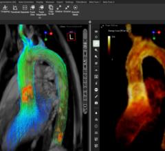
The Siemens Healthcare syngo.MR Cardiac 4-D ventricular function software calculates relevant parameters and automatically segments the right ventricle. MRI analysis software help automate segmentation and calculations to more quickly assess patients.
Increasingly software applications are providing many of the major advancements in magnetic resonance imaging (MRI) rather than hardware. In recent years, this has translated into software that enables physicians to do more in less time, extract more diagnostic information from imaging exams and provide more accurate quantification. While a hospital may not spend millions to upgrade its MRI system, it frequently takes advantage of the lower cost option of other vendors’ software to assess and manipulate those images.
A considerable amount of time can be spent slicing dataset images and using filters to find the exact part of the anatomy and information needed. This has been a barrier to wider adoption of MRI advanced visualization analysis software, but automation has drastically reduced the time needed manipulating images.
In addition to being the modality of choice for anatomical soft tissue imaging, MRI also offers functional assessment, which is enabled by newer software.
Analysis
Today’s MRI analysis software can calculate ventricular volumes, ejection fraction, myocardial mass and myocardial thickening for all segments. Many times these functions are automated to save time. Some software can perform dynamic evaluations, such as time-volume diagrams, filling rate and myocardial wall motion. It also presents information and a graphical display of results in tables, bull’s-eye plots and 4-D models of the heart. In addition, the information obtained by the software has the ability to be integrated into radiology or cardiology reports for documentation.
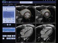
TomTec introduced its new 2-D Cardiac Performance Analysis MR software product during the Society for Cardiovascular Magnetic Resonance (SCMR) 2012 meeting. The tool quantifies myocardial function and deformation based on regular cardiac MRI. It allows a cardiologist to study the function of the heart muscle quickly and easily, offering quantification of displacement, velocity and strain in individual muscle segments to help assess hypertrophic cardiomyopathy or dyssynchrony of ventricles.
2-D Cardiac Performance Analysis MR is a feature tracking-based analysis tool that supports 2-D data from various cardiac MR systems. It analyzes myocardial deformation in standard MR cine sequences and does not require specific acquisition modes like tagged imaging. Existing patient data can be used for follow-up comparisons.
The software, which gained FDA approval in April, is a vendor independent software solution for the quantification of myocardial function with “cardiac MR cine images”. It presents a novel method to analyze myocardial deformation from routine cardiac MR images, which have been acquired as part of a conventional cardiovascular MR examination – no time-consuming tagging
is necessary.
Access
TeraRecon launched its iNtuition Review, iNtuition Enterprise Medical Viewer (iEMV) and iNtuition Share software enhancements in September, which enable Web-based access to multimodality advanced visualization software. It also allows easy sharing of images and datasets over the Web.
Automation
All MRI software vendors have automated features of their software so when a pre-specified type of study is opened, it renders “hands-off” in a pre-processed format without the need for user manipulation. Standard on some systems is auto segmentation of the coronary arteries and the endo- and epicardial left ventricular heart wall on short- and/or long axis images.
An example of automation using Siemens’ syngo.MR Cardiac 4-D Ventricular Function software is instant display of preprocessed segmentation of the left ventricle without user intervention. The software automatically calculates ventricular volumes, ejection fraction, myocardial mass and myocardial thickening for all segments. It also has semiautomatic processing of the right ventricle.
Web-Enabled Analysis
Calgary Scientific received U.S. Food and Drug Administration (FDA) clearance to market earlier this year for its ResolutionMD Web 2.9 Vessel Analysis module for post-processing diagnostic review and analysis of patient computed tomography (CT) and magnetic resonance imaging (MRI) scans. The module allows physicians, technologists and surgeons to review, edit, analyze and report findings of vascular anatomy across applicable specialties. Having the ability to use these tools over the Web brings added value as physicians can access images from home computers and laptops, providing freedom to work anywhere, even when an advanced clinical application or analysis is needed.
Beyond the Coronaries
Specialized software is available for peripheral and carotid artery and neurovascular imaging.
GE Healthcare recently partnered with VPDiagnostics Inc. to co-market the VPDiagnostics’ MRI-PlaqueView software for carotid artery atherosclerosis analysis. The software is compatible with carotid magnetic resonance imaging (MRI) data acquired from GE 3.0T MRI systems. The collaboration aims to develop optimized carotid imaging protocols and future co-developments on new techniques. Carotid atherosclerosis may be responsible for as much as one-third of all strokes and the software will help assess the risk of stroke from carotid atherosclerosis
VasSol’s Noninvasive Optimum Vessel Analysis (NOVA) technology provides detailed, quantitative information of blood flow through any vessel in the brain. Information provided includes velocity, volume and direction. It provides important quantitative data on what’s happening inside a blood vessel, as well as enhanced vessel visualization that raw MRI alone can not provide. The software does not require contrast injections.
Comparison Chart
This article served as an introduction to an MRI software chart comparing verious vendor's software. To access the chart, click on the "comparison charts" link on the navigation bar at the top of the page. Companies that participated in the chart include:
aycan Medical — www.aycan.com
AZE Ltd. — www.aze.co.jp/en
Circle Cardiovascular Imaging Inc. — www.circlecvi.com
Diagnosoft — www.diagnosoft.com
Fujifilm Medical Systems — 3dimaging.fujimed.com
Pie Medical Imaging — www.piemedical.com
Siemens Healthcare — usa.siemens.com/syngovia
TeraRecon — www.terarecon.com
Vital Images Inc. — www.vitalimages.com

 January 21, 2026