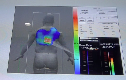
Toshiba is developing a radiation dose alert to show interventionalists how much dose they have delivered to their patient from X-ray angiography.
The latest advances in cardiovascular imaging are usually shown first at the Radiological Society of North America (RSNA) annual meeting, the largest radiology show in the world, held the last week of November in Chicago. After spending five days walking three expo halls filled with more than 600 product vendors, the following is my editor’s choice for the most innovative new cardiovascular imaging technology.
New Angiography Systems
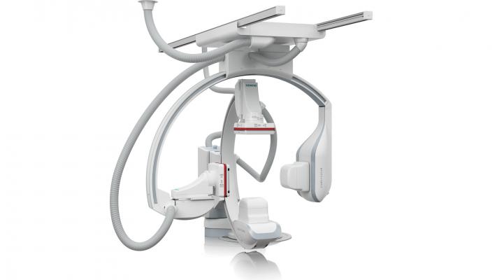
Siemens unveiled two new 510(k)-pending angiography systems, the Artis Q and Artis Q.zen, which incorporate new X-ray tube, detector and imaging software technology that can help reduce dose significantly, while offering improved image quality.
The new X-ray tube is intended to help physicians identify small vessels up to 70 percent better than conventional X-ray tube technology. The Artis Q.zen combines this innovative X-ray source with a new detector technology designed to support interventional imaging in ultra low-dose ranges to patients, doctors and medical staff, particularly during more complex, longer interventions.
The second generation of Siemens’ flat emitter technology replaced the coiled filaments used in conventional X-ray tubes to emit electrons. Flat emitters are designed to enable smaller quadratic focal spots that lead to improved visibility of small vessels.
The Artis Q.zen combines the X-ray tube with a detector technology that allows detection at ultra-low radiation levels. It can image with doses as low as half the standard levels applied in angiography. Instead of detectors based on amorphous silicon, a new crystalline silicon structure of the Artis Q.zen detector is designed to be more homogenous, allowing for more effective amplification of the signal, greatly reducing the electronic noise.
Siemens also introduced new software applications for interventional imaging. Clear Stent Live freezes an enhanced image of a stent during deployment with the balloon radio-opaque markers and uses it as an overlay on live fluoroscopy. Siemens says the main application will be for better visualization when implanting overlapping stents or stenting bifurcation lesions. It also helps suppress and stabilize heart motion on the image.
Other new 3-D applications are designed to image the smallest structures inside the head. Their high spatial resolution is crucial for imaging intracranial stents or other miniscule structures such as the cochlea in the inner ear. Moving organs such as the lungs can be imaged in 3-D in less than three seconds, reducing motion artifacts and the required amount of contrast agent.
GE Healthcare showcased its IGS (Image Guided System) 750 hybrid OR angiography system. It was displayed at RSNA 2011, but did not receive FDA clearance until earlier this year. It offers the mobility of a mobile C-arm, but the image quality and software features of a ceiling or floor mounted fixed system. It uses laser guidance for very accurate positioning. It can rove around the room on a powered caster system to enable different positioning around the table, or be parked out of the way during open surgical procedures.
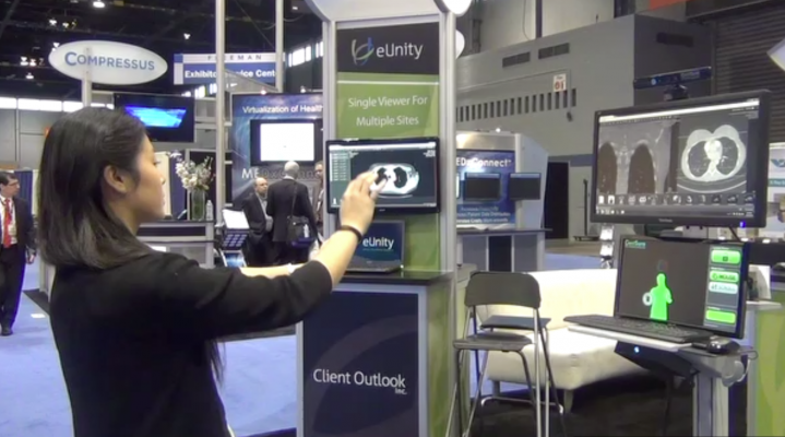
Hands-Free Physician Control of Images
GestSure displayed a new, FDA-cleared system that allows interventionalists in the cath lab, or surgeons in the operating room, to pick reference images to display on the overhead screens in the room and manipulate the images all hands-free. It allows physicians to pick and enlarge the images they need for better procedural navigation, while maintaining the sterile field.
A video sensor detects all the people in the work area and displays their outlines on a separate screen, with each person assigned a specific color. When one of those people raises their arms in the “hands up” pose, the system detects this and allows the person control of the system. Using the right arm/hand, they can scroll through images and use the left arm/hand as a mouse click by a pushing motion forward. The system detects the motions and translates them in real time to mouse actions on the overhead screen.
The software works as a vendor-neutral layer on top of existing PACS or advanced visualization software.
Outpatient, Office-Based Catheter Interventions
Outpatient, office-based peripheral vascular procedures are an increasing trend, according to GE healthcare, which showcased a new “mobile hybrid OR” solution. The trend includes setting up an outpatient cath lab in an office setting to reduce the costs of using hospital ORs or cath labs. The room system GE highlighted centers around its OEC 9900 Elite mobile C-arm and Venue 40, which is combined with a ultrasound system in an all-in-one unit. The GE Venue 40 tablet ultrasound system is mounted within the OEC 9900 Elite C-arm’s workstation to reduce the floor space required.
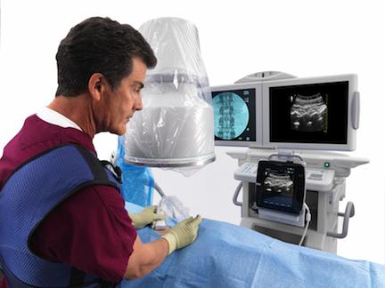
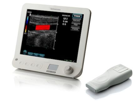
Wireless Ultrasound Transducer
Siemens introduced the world’s first wireless transducer ultrasound system, the Acuson Freestyle. It eliminates the impediment of cables in ultrasound imaging by using a battery-powered transducer, about the size of a large TV controller. The transducer can be submerged for cleaning. It is capable of 90 minutes of continuous scanning before the battery needs to be recharged.
The Freestyle is a point-of-care system that will expand ultrasound’s use in interventional and therapeutic applications. The transducer can be used to image up to 10 feet from the console. Siemens said it hopes to refine and expand the wireless transducer technology to its other systems in the coming years.
Engineers had to overcome several issues to create a wireless transducer. For example, a cardiac echo requires about 40 frames per second and each frame is equal to about 1 megabyte of data. To accommodate the amount of data and speed the computer processing involved, some of the electronics are placed in the transducer rather than processing the data in the machine console. The wireless system transmits the data over an 8 GHz ultrawideband radio frequency to the console. The amount of data and the bandwidth transmitted by the transducer is equal to about 10 4G smart phones working continuously.
Noiseless MRI
GE Healthcare introduced its 510(k)-pending noiseless MRI Silent Scan technology that it hopes to introduce in 2013 for its MR450W 1.5T system. The technology addresses one of the most significant impediments to patient comfort — excessive noise generated during the exam that can be in excess of 110 decibels. A combination of software and a pulse sequence lowers the noise level to that of a chirping bird outside a window.
Historically, acoustic noise mitigation techniques have focused on insulating components and muffling sound as opposed to treating the noise at the source. With Silent Scan, acoustic noise is essentially eliminated by employing a new advanced 3-D acquisition and reconstruction technique called Silenz, in combination with GE Healthcare’s proprietary design of the high-fidelity MR gradient and RF system electronics. Silent Scan is designed to eliminate the noise at its source.
640-Slice CT Scanner
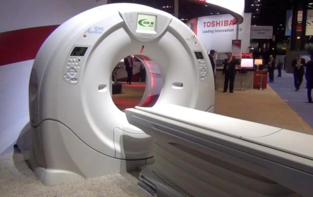
Toshiba unveiled its 640-slice Aquilion One Vision edition CT scanner. The vendor already offers the highest-slice system on the market, the 320-slice Aquilion One. The new system is equipped with a gantry rotation of 0.275 seconds, a 100 kw generator and 320 detector rows (640 unique slices) covering 16 cm in a single rotation, with the industry’s thinnest slices at 500 microns (0.5 mm). The system can accommodate larger patients with its 78 cm bore and fast rotation, including bariatric and patients with high heart rates.
FFR-Like CT Culprit Vessel Analysis
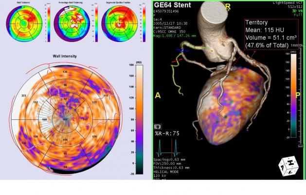
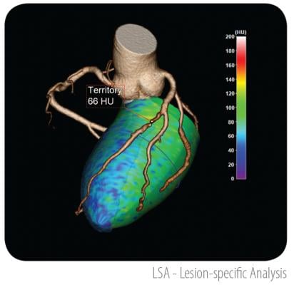
TeraRecon released new research software in response to fractional flow reserve (FFR)-CT analysis being developed by HeartFlow. The HeartFlow software uses a supercomputing algorithm to look at the fluid dynamics of the iodine contrast flow in coronary vessels to calculate a virtual a FFR number, similar to invasive pressure wire based FFR in the cath lab. TeraRecon’s Lesion Specific Analysis software cannot calculate FFR, but uses the same principle of tracking contrast flow in the myocardium. It uses lobular decomposition to look at each vessel segment to determine the tissue it feeds to show areas of ischemia and the expected culprit vessel segment. It shows a color contrast level maps on a 3-D model of the heart and in a coronal view of the left ventricle. Automated detection boxes highlight suspected ischemic areas of interest and identifies the vessel responsible for supplying blood to the region.
Radiation Dose Monitoring
Radiation dose monitoring solutions have been shown at previous RSNAs, but were highlighted by several companies this year as several states began implementing requirements for radiology departments to record patient dose. Dose records will have the most application with CT systems, especially for longer duration, higher dose cardiac exams, and catheter based angiography. Angiography is becoming an increasing issue due to the longer duration of more complex transcatheter interventions.
Toshiba demonstrated a work-in-progress dose tracking software for its Infinix-i angiography system. It can be displayed on a screen in the cath lab to show the approximate radiation dose that has been delivered cumulatively to specific areas of a patient. It takes into consideration the amount of time, power setting used and orientation of the C-arm to show a color-coded map of radiation delivery projected on a human figure. The colors change in real time as X-ray imaging continues. It is designed to be a visual reminder to physicians about the dose the patient has received and that they may want to change the location of the C-arm.
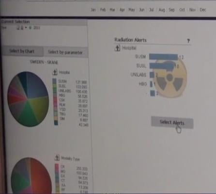
Sectra demonstrated 510(k)-pending Dose Track software, which radiology or cardiology departments can use to track radiation dose by patient, machine, physician, technologist, procedure type and room. The system can be set up to create alerts if a reasonable amount of dose if exceeded for a particular exam, or if certain physicians or technologists are using higher than average doses.
OLED Displays
Flat panel display technology migrated from CRT screens to LCDs over the past decade. The next major innovation in display technology is OLED, which offers even smaller components, faster response time than LCD, and the ability to display quick motion with virtually no blur. Sony showed the new PVM-2551MD OLED medical-grade monitor, which incorporates technology to achieve pure black, faithful to the source signal. By providing superb color reproduction, especially for dark images, surgeons can observe very subtle details such as the faint color difference between various tissues and blood vessels.
Aesthetically Pleasing Cath Labs
Philips Healthcare displayed video of its recent install of the Ambient Experience in a cath lab. The system uses colored lighting, subtle room design details and projected image visual effects to calm patients and make procedure rooms look less clinical. The installation highlighted allowed doctors or patients to choose a theme, such as a tropical rainforest, where diffused, indirect lighting would take a green hue and a photo projection on the ceiling of a tropical scene. Philips said at facilities that have installed these type of labs, patient satisfaction rose, as did staff morale. They say doctors and staff compete to use these rooms at some facilities.
Single Detector Spectral CT Imaging
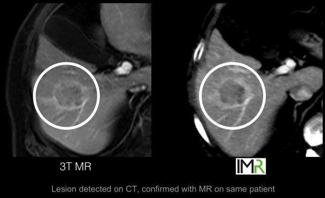
Philips introduced an innovative work-in-progress CT system that uses new detector technology to simplify spectral imaging, offering soft tissue image quality similar to MRI. Currently, CT special imaging can be performed using systems with two X-ray tubes and two detectors. The new system in development uses a single X-ray source and a single detector that has two layers of detectors, one on top of the other, for high and low energy.
Better Transcatheter Mitral Valve Repair Guidance
Philips’ showed its new Echo Navigator system, designed to synchronize views from TEE ultrasound with the orientation on live angiography. The primary application is to aid navigation during transcatheter mitral valve procedures, which require very accurate 3-D echo navigation to deploy devices like the Abbott MitraClip.
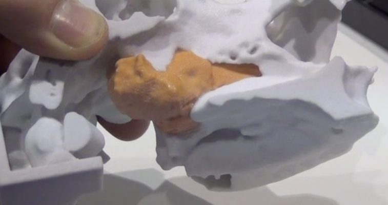
3-D Sculptures From 3-D Datasets
Taking 3-D images shown on 2-D display screens to a true physical 3-D form, Vidar Systems/3D Systems displayed the new Z Printer 450. It takes any 3-D advanced visualization dataset and can print the image in true 3-D using gypsum powder (the same material used to make drywall), standard color ink jet printer cartridges and a binding agent. The image is saved as an STL file and sent to the printer, which prints 1/10th of a millimeter each pass, up to 2 cm per hour.
The 3-D sculptures it created can be printed in color, eliminating the need to paint the models.
The printer offers a new way to create 3-D anatomical models for medical education, complex surgical planning and cosmetic reconstruction. Another application suggested at RSNA was to print sculptures for sale to the patients, such as fetal faces taken from 3-D obstetrics ultrasound exams.
The company printed a full-sized, 3-D, color heart during the show using a cardiac CT dataset on a thumb drive provided by one of the advanced visualization vendors in the same hall.
Video: New Cardiovascular Imaging Technologies at RSNA 2012
To watch a video version of this story, visit http://bit.ly/UIxYod.


 February 02, 2026