An example of radiation dose tracking software from Imalogix showing data on cath lab angiography cases. Photo by Dave Fornell
More than a decade ago, there was an alarming, rapid rise in ionizing radiation exposure in the U.S. population that was mainly driven the increased use of medical imaging. One of the biggest drivers was the rapid expansion of computed tomography (CT). However, radiology society driven campaigns to lower doses and new technologies developed by imaging systems vendors have succeeded in cutting radiation exposure rates in the most recent assessment.
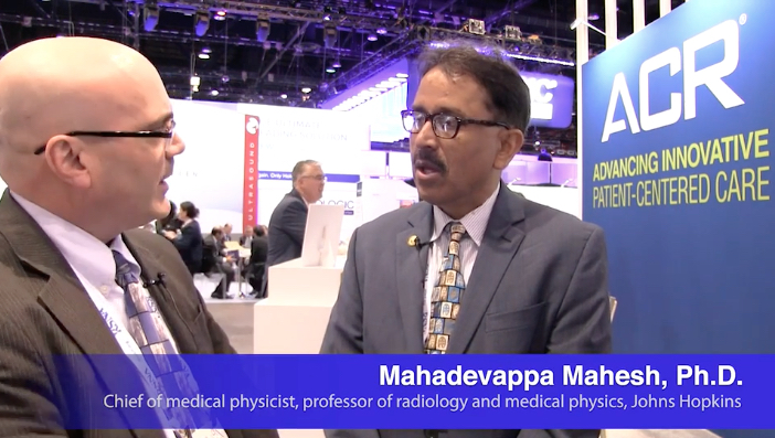
“We had a big report in 2009 called the Radiation Exposure to the U.S. Population, and it showed a dramatic rise medical radiation exposure. But, this new report shows that the overall radiation exposure to the U.S. population has gone down by between 15-20 percent over the past 10 years,” explained study co-author Mahadevappa Mahesh, Ph.D., chief of medical physicist and professor of radiology and medical physics, Johns Hopkins University, Baltimore, treasurer of the American Association of Physicists in Medicine (AAPM), a board member of the American College of Radiology (ACR). He is also the co-chair of the National Council on Radiation Protection and Measures Report (NCRP), and presented the most recent NCRP data analysis at the 2019 Radiological Society of North America (RSNA) meeting.
The NCRP 184 report covers the period between 2006 and 2016, the period of the most current Centers for Medicare and Medicaid Services (CMS) data. It showed sizable decreases in the radiation dose exposure in the U.S. population from medical imaging, compared to the 2009 NCRP 160 report, which covered the period prior to 2006.
Key findings of the study include:
• CT dose dropped about 6 percent, despite a 20 percent increase CT scans since 2006;
• Drop of more than 50 percent for nuclear imaging scans, mainly due to fewer procedures begin performed;
• A 15-40 percent decrease across all X-ray imaging modalities.
Decreases show the impact of using "as low as reasonably achievable” (ALARA) principals, new dose guidelines outlined jointly by numerous medical societies, and American College of Radiology (ACR) dose reduction initiatives like Image Wisely, Image Gently, and the ACR Dose Index Registry, Mahesh said
Mahesh, and many other medical physicist and radiology experts, have called for wider use of radiation dose monitoring software, because a hospital or imaging center needs to establish a baseline for its use of dose before they can measure the impact of any dose lowering efforts, changes in protocols, use of newer imaging equipment, or use of dose-lowering techniques. Some hospitals adopted dose recording software to improve their imaging protocols to improve safety for their patients and staff, but it was new recording requirements by the Joint Commission and some states that helped push many other facilities to adopt the technology.
He said there was growing concern a decade ago when the last council report was published, which showed a steep increase in radiation dose. This was mainly due to a rapid increase in the use of CT and other types of X-ray based and nuclear radiotracer medical imaging. This prompted the ACR to create the Image Wisely program and push for the use of more thoughtful imaging doses based on patient size, using the ALARA principle.
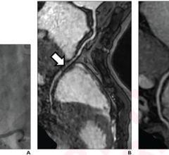
“The average dose to patients has gone down by 6 percent while CT exams have increased about 20 percent and we added new procedures like cardiac CT and CT perfusion,” Mahesh said. “Cardiac CT was one of the prime examples where we really saw the dose go down. In 2006, we were doing cardiac CT angiography exams with about 15-20 mSv of dose, but with the use of prospective gating CT technology it went down to 7-8 mSv. Then we had CT dose modulation, where we can do it at 3-5 mSv. If you are really enthusiastic and work at it, you can now go below 1 mSv. We have been able to do this in a decade and without the loss of image quality.”
Declines were seen across all the imaging modalities. Cardiac interventional angiography dropped 43 percent, non-cardiac interventional angiography dropped 40 percent, and radiography and fluoroscopy dropped 27 percent. However, he said the biggest decline was in nuclear imaging, where there was a 56 percent drop in dose. This was mainly driven by a decreasing number of nuclear exams, with many exams now being performed by CT.
“The technological advances over the past 10 years are really having an impact,” Mahesh explained. “For example, digital detectors are a lot more dose efficient so we don’t need the same amount of radiation to get the same image quality.”
Other technologies that have had a big impact on lowering dose include CT dose modulation, iterative reconstruction to improve image quality of lower dose CT scans, and dose recording and reporting software. “Now we have manufactures giving CT dose notifications so the users know when they exceed certain dose thresholds,” Mahesh said.
Dose recording software has helped prevent “dose creep” by the technologists who slowly increase dose of the imaging systems because it results in improved image quality. But now, systems send alerts when protocol limits are exceeded and can track which staff members are not following ALARA principles. This software also has helped lower the amount of dose received by staff.
“Also, over the past 10 years, we seen a lot more increased awareness about radiation dose and optimization,” he added. “If you are aware of the doses you are using, you will try to be more careful about optimizing the protocols. We also now have accreditation requirements to record your doses. Accreditation allows the sites to look at their protocols. Not that this is going to solve all the problems, but at least it’s giving a look at dose and can consider optimizing.”
He said participating in the ACR Dose Index Registry also provides hospitals feedback on what other sites are doing regarding dose and how your facility compares, which is a motivator to lower dose.
“We are also seeing the staff radiation exposure decline in X-ray and other places, because as we lower the patient dose, everybody benefits,” Mahesh said.
Find out more from this study in a VIDEO interview with Mahesh.
Related Medical Imaging Radiation Dose Resources:
VIDEO: Radiation Dose Monitoring in Medical Imaging — an interview with Mahadevappa Mahesh, Ph.D.
The Basics of Radiation Dose Monitoring in Medical Imaging
Radiation Dose Monitoring Product Comparison Chart
How to Understand and Communicate Radiation Risk — Image Wisely
Radiation in Medicine: Medical Imaging Procedures
FDA White Paper: Initiative to Reduce Unnecessary Radiation Exposure from Medical Imaging
Radiation Dose in X-Ray and CT Exams
Radiation Dose from Medical Imaging: A Primer for Emergency Physicians
Radiation risk from medical imaging

 October 24, 2025