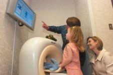
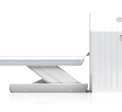
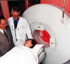
Philips Ambient Experience gives patients control over their surroundings as well as providing a soothing distraction.
According to the Centers for Disease Control and Prevention, more than 70 million Americans currently live with cardiovascular disease, with 700,000 dying from it each year.
In an age where heart disease is responsible for 29 percent of all U.S. deaths, cutting-edge technology and know-how is crucial to today’s cath labs.
To provide patients with the latest, most effective diagnosis and treatment for cardiovascular diseases and conditions, clinicians need access to state-of-the-art technologies in imaging modalities, patient monitoring and information management and customizable environments.
Imaging modalities shift toward CT, hybrids
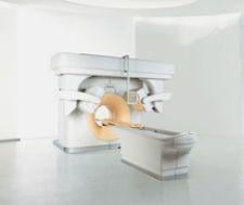
Heading up today’s cutting-edge cath lab are advanced imaging modalities, capable of providing dramatically clear, quality images and consistency. With modalities such as CT and hybrid imagers beginning to take centerstage, throughput time is going down and diagnosis is happening earlier and faster.
“I think it’s just natural that CT is being used more in the cath lab,” said William Guy Weigold M.D., director of Cardiac CT at Washington Hospital Center. “The CT angiograms produce such beautiful, exquisite images of the coronary vessels and also do such a good job of detecting, pinpointing and assessing disease severity.”
According to Dr. Weigold, having a CT in the cath lab saves time and money and is safer for the patient, who may not have to go through as much radiation if a diagnosis can be made sooner.
“Rather than simply generating a report that says there is a lesion in this part of the coronary vessel and then having an operator in the [cath] lab have to go and redo everything over again as far as diagnosis, it just makes sense to bring that information into the cath lab and use it in a detailed way in which the operators are looking at the CT angiogram and the actual images themselves and even manipulating the data in three dimensional space, in the lab, in order to save time, contrast and radiation,” said Dr. Weigold, who uses Philips’ Brilliance CT 64-slice scanner with Extended Brilliance Workspace in his cath lab.
Philips’ Step & Shoot Cardiac is the newest cardiac imaging innovation for the Brilliance CT 64-slice configuration. This application provides high-quality CT images of the coronary arteries and heart anatomy at low radiation dose levels and short breath holds.
“We have particularly widespread interest in using CT angiography in a lot of patients and are very sensitive to the fact that radiation is involved,” said Samuel Wann, M.D., chairman of Cardiovascular Medicine at the Wisconsin Heart Hospital. “It’s good to be wise and use as low a dose as possible.”
According to Dr. Wann, Step & Shoot uses around 2-3 millisieverts, whereas conventional techniques supply a radiation dose of 12-14 millisieverts. Such a drop in radiation level reduces any major concerns about the dose, even in younger patients.
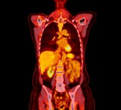
Hybrid imaging modalities such as PET/CT, SPECT/CT and the up- and-coming MR/PET are also being welcomed into the cath lab with open arms. These hybrids combine commonly used modalities to form a one-stop-shop for cardiac care.
Hybrids such as Philips’ Precedence SPECT/CT provide comprehensive cardiac imaging in a single episode in order to display anatomical localization as well as metabolic function.
Though it is still in development, MR/PET has the potential to provide soft tissue content as well as simultaneous data collection. Because a patient’s condition can change even five seconds after an image is taken, being able to perform PET and MR at the same time has its benefits.
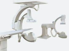
Other innovations in imaging modalities include Toshiba’s Infinix CF-i/BP with five-axis positioner, which allows unrestricted patient access from virtually any angle and fingertip-to-fingertip and head-to-toe coverage for cardiac imaging, according to Toshiba. The system allows clinicians to obtain bi-plane views of any region of the heart with a C-arm design that provides reportedly unparallel access to the patient in addition to 8-inch flat panel detectors for distortion-free images.
According to Dr. Wann, CTA images can be co-registered on such modalities with the C-arm to help select the best angles and to measure the best length for stents.
Data review, regardless of location
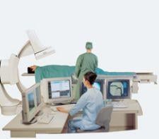
Patient monitoring no longer consists of having a person “plugged-in” to a machine and then into the wall. New cath lab technology requires adherence to the wireless, and paperless, world.
Devices such as the Philips HeartStart MRx Monitor/Defibrillator connect to a hospital’s real-time clinical network and allow for wireless transport. Patient data is transmitted via Internet to a receiving station, whether in the department itself or to a cardiologist's PDA, giving the physician complete access to patient data, wherever they are.
According to Scott Cochran, manager of Cardiac and Imaging Services at Carson Tahoe Regional Healthcare, one interesting new aspect of patient monitoring centers is the amount of data being collected with each procedure.
“In the past we were just monitoring the procedure and the hemodynamics and giving the physicians a report about that, but in today’s world, it’s all about inventory management and outcome data,” said Cochran.
Information management solutions allow for an integrated workflow and add to the quality and efficiency of patient care, all while collecting and displaying patient and image data wherever it is needed, with the push of a button.
“The newest versions of [Philips’] Xcelera allows us to look at coronary echoes, nuclear images and CTA images on the same workstation,” said Dr. Wann.
Personalized environments ease patient stress
When highlighting cutting-edge cath lab technology it is easy to overlook the most important aspect of any cardiovascular procedure: patients and physicians. The cold tables, glaring lights and frightening labs of the past are now yielding to a friendly and focused environment, making both patient and staff more comfortable in the cath lab.
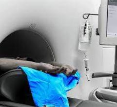
Philips’ Ambient Experience addresses many environmental concerns for both the patient and the cath lab staff. Ambient Experience allows patients to select a theme upon entering the lab. Lights are dimmed and a scene, such as an island beach or desert dunes, is projected onto the wall, allowing a positive distraction that is beneficial for patients of all ages.
Today’s comforting cath labs include warm tables, subdued lighting and a de-cluttered space to calm patients’ nerves.
“We like to keep the patient warm and informed in order to help them relax,” said Cochran. His associate, Annette Patellos, director of Cardiac and Critical Care Services agrees. “We also try to keep our lights as dim as possible and use heated blankets and contrasts,” she stated.
According to Kristin Cusolito, director for Ambient Experience for North America, by allowing patients some level of control, such as choosing the room’s theme and allowing them privacy when changing and during certain procedures, the process is easier for all involved.
“In this environment, the patient aspect is important, but even more so is the staff and the comfort of the staff,” said Cusolito. “Certain elements of the workflow, architectural design, de-cluttering of the workspace, storage cabinets and how they are all laid out really make a difference.”
Seemingly simple tasks, such as positioning lighting so that no reflections show on monitor screens can have a great effect on staff, according to Cusolito.

 November 17, 2025


![Phase III clinical trial of [18F]flurpiridaz PET diagnostic radiopharmaceutical meets co-primary endpoints for detecting Coronary Artery Disease (CAD)](/sites/default/files/styles/content_feed_medium/public/Screen%20Shot%202022-09-13%20at%203.30.13%20PM.png?itok=2w6OoNd6)