June 12, 2014 — Ischemic heart disease, a narrowing of the arteries supplying blood to the heart, is a leading cause of death throughout the world. A hybrid molecular imaging technique called positron emission tomography and magnetic resonance imaging (PET/MR), which tells doctors vital information about cardiac and arterial function, has been found to be an effective molecular imaging tool for detecting coronary artery disease (CAD), say researchers at the Society of Nuclear Medicine and Molecular Imaging’s 2014 Annual Meeting (SNMMI).
Often patients suspected of having CAD undergo a stress test called myocardial perfusion imaging (MPI) to smoke out areas of arterial ischemia and risk of myocardial infarction, or heart attack. Many undergo a molecular imaging scan called single photon emission computed tomography (SPECT), but in recent years PET/MR has emerged as a potential alternative due to its superiority for imaging the structures of soft tissues and the physiological function of the heart. This clinical study showed that PET/MR is consistently accurate using coronary angiography as the reference standard for detecting CAD.
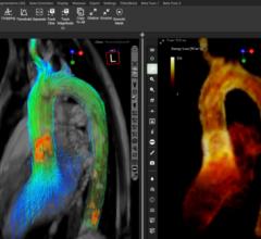
“By combining two advanced imaging modalities, PET and MR, cardiac PET/MR imaging allows a union of anatomic information with MR and functional information with PET for a comprehensive view of the of the heart,” said principal author Jeffrey M.C. Lau, M.D., Ph.D., from Washington University in St. Louis, Mo. “This allows us to predict or rule out coronary artery disease with more certainty, and in some instances, it allows us to detect disease processes such as areas of hibernating heart muscle that would not have been detected using conventional stress testing methods like SPECT.”
In addition, cardiac perfusion PET/MR can be performed in a shorter timeframe than SPECT and is associated with a lower dose of radiation per procedure, and MR can be used to produce an almost of disease in the arteries of the heart when compared to industry standard cinematic, multiple-frame sequence of the motion in specific regions of the heart muscle, most notably the left ventricle, which pumps oxygenated blood back into the body through the aorta. PET also provides quantitative data about blood flow in addition to the visual interpretation of disease.
The study involved 10 patients with reversible ischemia as indicated by SPECT-MPI. Scientists administered a radionuclide PET imaging agent called N-13 ammonia plus an MR contrast agent called gadolinium and the pharmaceutical Regadenoson, which imitates the stress of exercise. The researchers optimized the cardiac PET/MR imaging protocol in order to register areas of reduced perfusion of blood using MR with PET data about myocardial blood flow. The results showed that PET/MR imaging was very accurate in diagnostic coronary artery diseases. In this small sample, PET/MR had 100 percent sensitivity, 80 percent specificity and 100 percent negative predictive value. Those numbers compare favorably to SPECT in this study group.
This study was conducted in conjunction with Siemens Medical Solutions and funded in part by Astellas Pharma Inc.
For more information: www.snmmi.org

 January 21, 2026