Healthcare institutions are trying to drive productivity by increasing patient throughput while still maintaining a high quality of care. The goal is usually to reducing imaging exam times to increase the number of patients seen each day. The biggest challenge may come from ultrasound, which has the highest degree of operator variability and reproducibility issues. The last few years, however, vendors in the space have introduced several new technologies and features to help achieve these timesavings.
Enhanced Image Quality, Acquisition
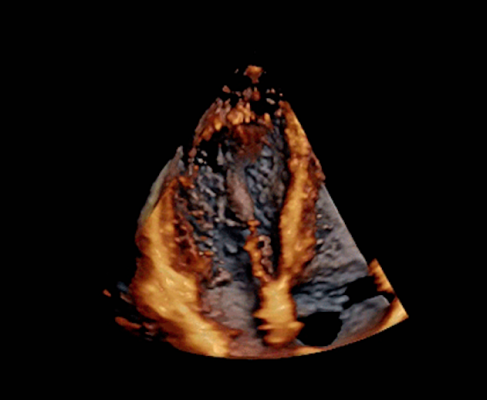
Any imaging modality is only as good as the image quality it can provide, and ultrasound manufacturers have been working to expand capabilities in this area. Three-dimensional and even 4-D ultrasound have been on the market for several years but are only now starting to increase traction in the marketplace as providers realize it can greatly reduce operator variability in acquisition and interpretation.
“It’s one of the most significant things you have to overcome,” said Jon Brubaker, MBA, RCVT, ultrasound market analyst for MDbuyline. “When you’re using 2-D, you need to do it on repeated cases, and if you’re not getting the exact image or location, it can affect what you’re seeing and ultimately your diagnosis.”
That has always been the goal of 3-D echocardiography, according to Federico Asch, M.D., associate director of the echocardiography core lab at Medstar Health Research Institute, Hyattsville, Md., and assistant professor of medicine (cardiology) at Georgetown University. “But it’s always been a theory; in practice we were not able to do that. But we’re getting there.”
One recent example of this is GE Healthcare’s new flagship ultrasound system, the Vivid E95, launched in the middle of 2015. The system offers 3-D and 4-D imaging, powered by GE’s cSound technology that captures and processes much larger amounts of image data than previous-generation systems — roughly a DVD’s worth of information per second. The algorithm also automatically removes image artifacts such as tissue coming in and out of plane.
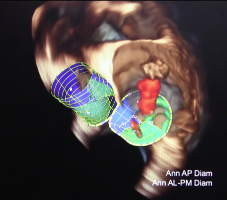
Siemens’ newest premium ultrasound system, the Acuson SC2000 Prime, performs real-time 3-D/4-D echo with live color flow Doppler overlaid on the image using a single-beat acquisition. The system also offers transesophageal echo (TEE) functionality, a technique seeing rapid growth in the face of dozens of new complex transcatheter structural heart device released in the last few years. TEE is perfectly suited for transcatheter aortic and mitral valve replacement (TAVR and TMVR) devices, left atrial appendage (LAA) occlusion and septal defect closures, which require high-precision, real-time imaging for device sizing, placement and real-time verification of closure or hemodynamic flow assessment.
The rise of 3-D and 4-D echo is not a signal, however, of the demise of 2-D ultrasound. Toshiba’s Aplio Platinum Series CV (cardiovascular) ultrasound systems, including the Aplio 300 Platinum CV and Aplio 500 Platinum CV, enhance image quality through a series of new features. Two-dimensional wall motion tracking, plus spectral and color Doppler, deliver high detail and resolution throughout the entire field-of-view, resulting in accurate and easy-to-use 2-D strain resolution.
Automated Data Quantification
Perhaps the most significant way that ultrasound exam times are being reduced is through automation of various processes, including data quantification. “Most of the vendors are trying to make the process of making measurements very streamlined and very easy so it doesn’t interrupt the workflow of any clinical lab, but at the same time provides an accuracy that is acceptable,” said Asch.
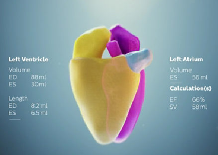
The 2015 American Society of Echocardiography (ASE) annual meeting saw the debut of HeartModel A.I., an advanced algorithm from Philips Healthcare delivering what the company calls Anatomically Intelligent Ultrasound (AIUS). First deployed on the Epiq 7 ultrasound system, HeartModel A.I. provides advanced quantification, automated 3-D views and robust reproducibility for cardiac ultrasound exams. The algorithm is built on a digital database of anatomical structural models, which are used to gather left ventricular and left atrial dimensions and volumes. These models automatically adapt to variability in patient anatomy, which the software has learned to do based on machine learning review of thousands of previously recorded echo exams.
Philips claims the technology is able to gather cardiac volume information three to six times faster than 2-D ultrasound systems. This allows physicians to more quickly and accurately assess disease states, determine treatment and guide related therapies. “I think that’s going to be really significant and important to the acceptance of it because it’s got to be able to process quickly, accurately and consistently,” said Brubaker. “Otherwise if it’s going to involve a lot of time and effort on the part of the people involved, acceptance is going to be hard. But I think it’s going to be pretty exciting.”
Siemens offers similar functionality through its eSie Valves software package, introduced on the Acuson SC2000 Prime in 2014. As the name suggests, this semi-automated advanced analysis program captures valve measurements during cardiac procedures. Where other systems may take several minutes to provide this information, eSie Valves returns aortic and mitral valve measurements in seconds.
While impressive, these early forays into artificial intelligence cannot yet offer automatic quantification of the entire heart. Asch highlighted the right ventricle as one example, but he believes the capability will one day be available. “At the end of the day it’s the same technology that needs different algorithms to be applied to a structure that has a completely different shape,” he told DAIC.
Touchscreen and Tablet Systems
As tablets and smartphones become everyday devices for a large majority of the population, many imaging system manufacturers are shifting their user interfaces to mirror mobile device functionality. In the ultrasound market, this has led to several new systems that are touch-screen operated, tablet-based or both. Rather than using dozens of keystrokes to zoom, activate functions and move objects, users can tap, pinch, drag and swipe their way through an examination.
“That’s an approach to the market for non-sonographers,” said Brubaker. “This is something they’re familiar with. It comes with a touchscreen so they can easily get the images and don’t have to spend a lot of time getting the end product they need.”
Mindray adopted tablet technology for ultrasound last year with the release of the TE7 system. Though still cart-based, the TE7 completely eliminates the traditional ultrasound keyboard, even offering one-touch image optimization. Cardiovascular-specific functions on the system include continuous wave (CW) Doppler and a TEE transducer.
Carestream Health entered the ultrasound market at the 2014 Radiological Society of North America (RSNA) annual meeting with the introduction of the Touch Prime and Touch Prime XE premium systems. In this case, the touchscreen offers an appearance and layout similar to traditional keyboards with the flexibility of a soft user interface with full smart-touch functionality. Etched marks on the glass screen help users quickly locate primary buttons without having to look away from the screen. Users also can select the appropriate transducer type with a single touch.
Many of these systems with mobile device functionality are targeted for use at the point of care, where conditions may be different in every scenario. For these cases, a number of these technologies were on display at RSNA 2015, including:
● The iViz handheld ultrasound system from Fujifilm Sonosite, cleared by the U.S. Food and Drug Administration (FDA) just before the show. The high-resolution, 7-inch display touchscreen offers a wide dynamic range and vibrant color flow images, with multiple imaging modes and exams available with the tap of a finger. In what the company calls an industry-first, the iViz offers full bi-directional connectivity with the electronic medical record (EMR), meaning users can both send reports and receive patient demographics; the latter functionality eliminates manual entry of patient information for a significant time savings.
● The Lumify app-based ultrasound system from Philips Healthcare received FDA approval in July 2015. Rather than operate from a dedicated handheld system, Lumify turns any compatible Android smart device into a display screen simply by plugging in the USB transducer. The transducer handles all of the image acquisition and processing and is controlled through the downloadable cloud-based Lumify app. The app is free to download, but the transducer is rented on a monthly subscription basis.

 January 28, 2026