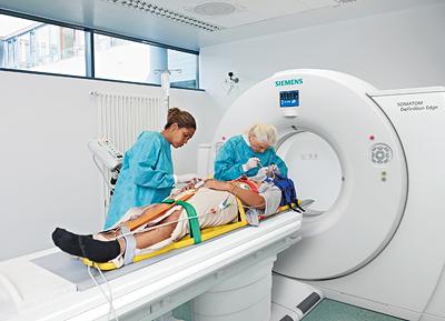
June 20, 2012 – Siemens Healthcare announced the Somatom Definition Edge single-source computed tomography (CT) system has been approved for sale in the United States. It is the first single-source CT to use Siemens’ recently introduced Stellar Detector.
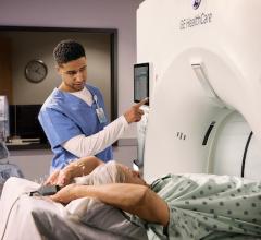
With ever-greater patient volumes in the emergency department (ED) setting, where 40 percent[1] of CT examinations are performed, hospital demand is increasing for more efficient and cost-effective imaging solutions. With these escalating ED needs in mind, Siemens designed the Somatom Definition Edge CT system.
The system’s Stellar Detector for the first time fully integrates the detector’s miniaturized electronics with the photodiode. It has the potential to significantly reduce electronic noise and cross-talk and improve the signal-to-noise ratio of the images. With the Stellar Detector, the Somatom Definition Edge can generate ultra-thin slices to deliver an extremely high spatial resolution with an optimal signal-to-noise ratio and without any increase in dose. The increased spatial resolution may potentially benefit critical cases such as emergency diagnostics and cardiovascular examinations, where every additional visible micrometer can be beneficial.
“Physicians are potentially able to visualize even the finest details of the vascular system or micro fractures of the bone, which may potentially provide more detailed information when they are attempting to identify coronary artery blockages or subtle spinal fractures that, if undiscovered, could result in paralysis,” said Kulin Hemani, U.S. vice president of CT, Siemens Healthcare. “These revolutionary innovations have the potential to help clinicians make more sound diagnoses, even for the most challenging cases such as trauma and bariatric patients. This is part of our continuing commitment to excellence in patient care, particularly in the ED environment.”
The Somatom Definition Edge CT system can enable visualization of structures up to 0.30 millimeters, with potential improvement in image sharpness. Siemens’ fastest single-source CT scanner, the Somatom Definition Edge has a rotation speed as fast as 0.28 seconds and can acquire up to 23 cm/s. And the new dual energy mode enables the system to scan at two different tube voltages to help better characterize tissue types.
Increased Speed in Single-Source CT
Maintaining high spatial resolution at high gantry rotation speeds has long proven challenging in CT. With patients who are unable to remain still for long periods, or with scans involving fast-moving organs such as the heart, the shortest possible scanning time may reduce image motion artifacts. The new gantry design of the Somatom Definition Edge CT system has a very high temporal resolution of 142 ms. The system allows a pitch of 1.7, thus enabling an acquisition speed of up to 23 cm/sec, and is capable of reconstructing 384 slices per rotation. With the scanner, a typical thorax-abdomen examination can be performed in about two seconds, potentially minimizing the need for patient breath-holding during scanning. A 6-foot patient can potentially be scanned in 8.3 seconds at full speed using the system.
Dual Energy for Single-Source CT
Until now, dual energy examinations with single-source devices were complicated processes that resulted in dose levels several times higher than levels seen with a customary single-source scan. The Somatom Definition Edge CT system introduces a new scanning technique into single-source CT examinations. During the dual energy examination, the machine performs two CT scans in succession with different energy levels, with each scan potentially using only about half the dose applied in normal mode. Together, the two scans deliver the image signal required. Also, since the voltage in each scan is fixed, the physician can use additional dose-reducing techniques, including CARE Dose4D – a fully automatic radiation exposure control system that adjusts the dose in real time according to the area of the body being scanned. Additionally, the Definition Edge uses the rapid iterative reconstruction method SAFIRE (Sinogram Affirmed Iterative Reconstruction), which may also help to reduce dose levels.[2] These Siemens offerings enable the generation of dual energy images via a single-source system without excessive patient radiation dose.
Advantages in Cardiovascular Imaging
The Definition Edge may also offer clinicians a major advantage with its marriage of high temporal resolution and the high degree of spatial resolution provided by the Stellar Detector. Since coronary arteries constantly move along with the heart, the clinician must obtain CT images at a high temporal resolution to sharpen coronary artery images. The sharpness of image provided may improve the clinician’s ability to detect coronary stenosis and atherosclerotic changes in plaque formations. And the high spatial resolution may potentially improve the physician’s ability to safely detect, for example, in-stent restenosis of stents smaller than 3 mm in diameter.
The Somatom Definition Edge CT system with Stellar Detector will begin shipping in the summer of 2012. The entire Somotom Definition AS and Definition Flash scanner platforms are upgradeable with the Stellar Detector.
For more information: www.siemens.com/healthcare
References:
1. IMV 2012 CT Market Outlook Report, May 2012.
2. In clinical practice, the use of SAFIRE may reduce CT patient dose depending on the clinical task, patient size, anatomical location, and clinical practice. A consultation with a radiologist and a physicist should be made to determine the appropriate dose to obtain diagnostic image quality for the particular clinical task. The following test method was used to determine a 54 to 60% dose reduction when using the SAFIRE reconstruction software. Noise, CT numbers, homogeneity, low-contrast resolution and high contrast resolution were assessed in a Gammex 438 phantom. Low dose data reconstructed with SAFIRE showed the same image quality compared to full dose data based on this test. Data on file.

 February 06, 2026