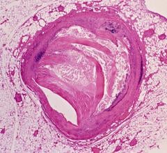
Angiographic imaging system vendors have developed several new technologies to address emerging cath lab trends, including the need to reduce radiation dose, improve image quality and enable advanced procedural image guidance. All three of these points have become increasingly important as more complex procedures are attempted in interventional cardiology cath labs and hybrid ORs. These procedures include transcatheter aortic valve replacement (TAVR), MitraClip repairs, left atrial appendage (LAA) occlusions, atrial and ventricular septal defect closures, and new interventions for both electrophysiology (EP) and heart failure.
All of the major vendors in the United States introduced new systems and technologies in the past few years to reduce dose and enhance visualization in the cath lab. The vendors have tailored their systems into various models for specific specialties and at various price points depending on the degree of functionality. Most vendors also offer software to enhance visualization of stents and other devices.
Advanced Imaging, Guidance, Planning
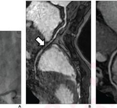
Newer imaging systems enable advanced 3-D imaging with rotational angiography, which uses a quick spin around the patient to create a computed tomography (CT)-like, 3-D image of the anatomy. This can all be done tableside in the cath lab. Some systems allow these images, or CT or magnetic resonance imaging (MRI) 3-D images, to be overlaid or fused with the live 2-D fluoroscopic images. This fusion technology is used with TAVR planning and navigation software to better guide precise device placement. Software also allows 3-D images to be integrated with EP electromapping systems to guide catheter ablation procedures without the need for live fluoroscopy, helping to reduce dose.
At the Radiological Society of North America (RSNA) 2014 annual meeting, Siemens introduced its Artis With Pure technology, which simplifies the use of Siemens' 3-D rotational angiography. The software suggests imaging protocols for ease of use and offers step-by-step instructions to create a fusion image.
Siemens' Syngo Dyna 4-D imaging now enables a combined digital subtraction angiography (DSA) and DynaCT (a CT-like 3-D image) at the same time to show filling of vessels with contrast. The Syngo DynaCT SMART feature can remove metal streak artifacts tableside from 3-D reconstructions. A Quick Zoom feature also allows zooming in on a region of interest tableside without the need to manually manipulate images in the control room. These technologies can be retrofitted on older Artis systems.
New software suites for various specialties have been introduced for Philips' angiography systems, including interventional oncology, interventional neurology and for the hybrid OR suite. The oncology suite can perform two passes with the C-arm for to collect images of the arterial and tumor phases with contrast to combine them into a single image to help guide embolizations. The software also features automated feeder vessel detection. Philips EchoNavigator software enables fusion imaging with live 3-D/4-D transesophageal echo (TEE), showing the relation of the TEE image on live or captured image angiography.
At RSNA 2015, GE showcased its Assist brand, a collection of new interventional imaging software packages designed for specific clinical subspecialists. The suite offers simple to use fusion imaging for computed tomography (CT) 3-D image reconstructions that can be overlaid or fused with live angiography in the cath lab. The new Assist suite of targeted software solutions include Vessel Assist to better navigate vessels for chronic total occlusions (CTO), transjugular intrahepatic portosystemic shunt (TIPS) and interventional neuroradiology arteriovenous malformation (AVM) and aneurysms. The EVAR Assist aids endovascular aortic repair (EVAR). Needle Assist is for bone interventions and pelvic bone osteosynthesis. FlightPlan for Liver helps with liver embolizations. Valve Assist is for transcatheter aortic valve replacement (TAVR). PCI Assist aids complex percutaneous coronary intervention (PCI) navigation.
At the 2015 American College of Cardiology (ACC) meeting, Toshiba highlighted its 3-D rotational angiography capabilities and its new transcatheter valve planning and image fusion capabilities. The 3mensio Structural Heart software facilitates pre-operative planning of aortic valve procedures, mitral valve procedures and left arterial appendage (LAA) closures. It can also fuse 3-D CT or MRI images with live fluoroscopy to enable procedural guidance.
Radiation Reduction
All the new angiography systems on the U.S. market offer dose-lowering technologies. These advances include improved X-ray tubes, more sensitive detectors and software to help improve image quality at lower doses, in addition to noise reduction. The Philips Allura Clarity is an example of the next-generation system that can help lower standard procedural dose by 50 to 75 percent. The operator can easily adjust the frame rates to help reduce dose, and the system incorporates Philips' ClarityIQ software to achieve excellent visibility at low X-ray dose levels for patients of all sizes. The software helps correct for motion, reduces noise, auto enhances the image and corrects pixel shift on cine images. The ClarityIQ technology is also available as an upgrade for the majority of Philips' installed base of monoplane and biplane interventional X-ray systems.
Toshiba's Spot Fluoroscopy software for its Infinix-i systems allows clinicians to observe a target region of anatomy using Spot Fluoro's live fluoroscopy, while viewing the last image hold (LIH) in the surrounding area that has been collimated out of the field of view to cut dose.
GE, Toshiba aqnd Philips offer software to visualize the dose accumulation (peek skin dose) on a patient in real time. All these systems show an illustration of the distribution of received X-ray radiation to serve as a reminder to move the C-arm to avoid radiation burns. Dose limits also can be set.
Newer Systems
Siemens introduced several new systems in its Artis series, including the Artis Q, Artis Q.zen and the Artis one. These incorporate both new X-ray tube and detector technology to not only lower dose, but also cut energy consumption by up to 20 percent from previous generation Artis systems. Siemens' new X-ray tube uses flat emitter technology, which enables smaller, square focal spots that the vendor said leads to as much as 70 percent improved visibility of small vessels and improved image quality. The Artis Q.zen combines this X-ray tube with a new crystalline detector that enables patient doses as low as half the standard angiography levels.
The Artis Q, Artis Q.zen and Artis one include the Clearstent Live application, which freezes motion in the region of a stent, allowing the physician to mask out movement of the beating heart and place the stent in precisely. The Artis one also offers new tools for cardiac imaging, including HeartSweep, which uses dual-axis rotational angiography to image the entire heart in a single, smooth C-arm movement instead of multiple acquisitions from different projections. This single-sweep movement has the potential to speed up procedures and save contrast agents.
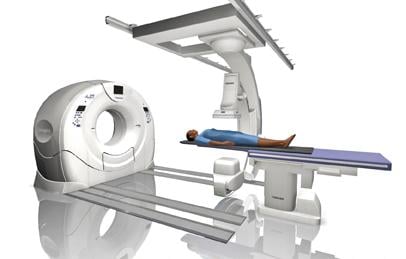
At RSNA 2014, Toshiba introduced its new line of combination computed tomography (CT)/angiography systems. It combines an Infinix Elite angiography system with an Aquilion Prime or Aquilion One Vision Edition CT system. This offers an all-in-one interventional lab and CT solution to deliver real-time CT images during procedures instead of CT-like rotational angiography images. It can significantly improve workflow with its SURE Guidance technology, allowing image transition between modalities. At RSNA 2015, Toshiba showcased its Infinix 4DCT, which merges the Infinix Elite angiography system and Aquilion One Vision edition CT system. The Aquilion One Vision is capable of capturing an entire organ in one rotation with 640 slices and 16 cm of anatomical coverage. The combination of the interventional lab and CT eliminates the need to transfer patients between departments and allows clinicians to decrease procedure time and maintain patient safety.
Shimadzu's new angiography system platform is the Trinias series. It uses high-speed Score Pro image processing technology, and a smart ergonomic design aids intuitive functionality. The Score process enhances fluoroscopic images while maintaining as low as achievable dose considerations.
Designed as an angiographic interventional system, Trinias offers a new 12-inch Crossover flat panel detector for multi-procedure versatility that accommodates any type of angiography procedure. Combined with Score Pro image processing, the systems provide excellent visibility and numerous image guidance functions, as well as sophisticated 3-D application techniques. When used as a cardiac or neuro interventional system, Trinias is equipped with a new 8-inch flat panel detector. The system also has Score StentView, which improves visibility of existing stents in vessels.
GE Healthcare introduced its revolutionary Discovery IGS series in 2013. The laser-guided system captures the advantages of floor- and ceiling-mounted fixed systems and the mobility advantage of mobile C-arms without any of their inherent limitations. The imaging device is on a mobile, wheeled gantry that can be moved anywhere in the room like a mobile C-arm, but offers precision, high-quality imaging and features of a fixed system. At the touch of a button, clinicians can move the system to the table for imaging various anatomies and then move it aside and park it, allowing room to optimally position physicians, nurses, technologists, anesthesiologists and other personnel for surgery, with unobstructed access to the patient. The gantry comes with a new wide-bore design, which allows for steep angles and ease in 3-D acquisition, especially for large patients. It uses the same digital flat-panel detector technology as GE's fixed-based Innova angiography imaging systems.
Detector Considerations
When looking at systems for labs utilized by multiple users (interventional cardiology, vascular surgeons, interventional radiology, etc.) the size of the X-ray detector should be kept in mind, as different users have different requirements. Detector size is expressed in one of two ways: height and width, or as a diagonal measure from corner to corner. So a 20 x 20 cm detector can also be described as a 10-inch diagonal detector.
Dedicated cardiac and neuro cath labs generally use smaller detectors in the range of 8-10 inch diagonal to allow steep C-arm angulations to get different views of the coronary anatomy. Larger detectors in the rage of 15-20 inch diagonal offer a larger field of view for peripheral procedures and interventional radiology.
Philips added a new 16-bit digital detector on its FD20 platform. This offers four times higher contrast and higher DQE. Also, the new detector is designed so it does not need active cooling.
Comparison Chart
This article served as an introduction for a comparison chart for angiography systems on the U.S. market. To access the chart, click on the "Comparison Charts" tab at the top of the screen. Participants in the chart include:
GE Healthcare
gehealthcare.com
Omega Medical Imaging
omegamedicalimaging.com
Philips Healthcare
usa.philips.com/healthcare
Siemens Healthcare
usa.healthcare.siemens.com
Toshiba
medical.toshiba.com

 October 24, 2025