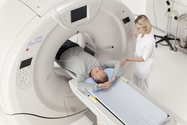
The Toshiba Aquilion One Vision system is among the new generation of CT systems on the market that offers several dose lowering technologies and a hardware/software combination to enhance image quality over previous-generation scanners.
As hospitals begin replacing their first-generation 64-slice computed tomography (CT) scanners after a decade of use, there are several considerations evaluation teams should think about when looking at the newer-generation scanners. Chief among these is the idea that more slices makes for a better scanner — which CT experts say is not necessarily the case. There are costs versus benefits to consider when looking at high-slice systems.
“The way you need to look at this is that the CT scanner is a tool, and you need the right tool for the right job, so it will depend on the hospital and what they plan to use it for,” said Claudio Smuclovisky, M.D., FACC, FSCCT, director of South Florida Imaging Cardiovascular Institute, Holy Cross Hospital, Ft. Lauderdale, Fla., and an expert in cardiovascular CT imaging systems.
“CT is really on the precipice and is really starting to accelerate in growth,” said Leslee Shaw, Ph.D., FACC, FASNC, FAHA, co-director of the Emory Clinical Cardiovascular Research Institute. “If you blink your eyes, you have missed several very prominent, randomized trials that support the utility of CT… We have more than just the pretty picture, we have seen dramatic growth in the technical aspects of CT with better image quality, better resolution, and we can go on and on.” She said all of these aspects are especially true for cardiovascular CT imaging.
Watch the video “What to Consider When Comparing 64-slice to Higher Slice CT Systems,” an interview with Claudio Smuclovisky at SCCT.
CT Coverage Area Versus Slices
Smuclovisky said there is a misunderstanding that more slices on a CT scanner means better images. He said a better measure is actually detector area coverage, which is the measurement of how much of the anatomy is being imaged at once. The more image area that can be covered determines if stitching several sets of images is needed to image an entire organ. This can lead to stitching artifacts and may require more time to reconstruct and review images, he said. This is especially true with movement caused by the heart or lungs.
Detector area coverage can vary between scanners with the same number of slices, because Smuclovisky explained the size of the detectors vary in size on each machine. In the case of 64-slice systems, he said they can range between 19.5 to 40 mm (4 cm) for detector area coverage. He said a system is considered a wide-area detector if it has 8 cm coverage or greater.
Wide detector systems tend to have a higher sensitivity, offer better iterative reconstruction software to improve both contrast and spatial resolutions, and they tend to have more powerful workstations, Smuclovisky explained.
“Most physicians don’t have detailed knowledge of the physics or the technology that is involved. So, a slice war started 10 years ago because people believed that if you had more slices, you would have better quality images," Smuclovisky said. "But, there are other components when you are doing high-end CT imaging. I tell people it is like looking at an airplane, where for the airplane to fly, it is not just how big the wings or fuselage are, it is the sum of all of its components. This includes the engines and the experience of the pilots — everything needs to fit together nicely and work together in a workflow so that plane can fly. So, it is not just about the slices, it is about many other components that go into a CT scanner.”
CT Rotation Speed
One key feature of scanners is the rotation speed of the gantry, which translates into faster temporal resolution to reduce motion blur, which is especially important with the heart and areas near the lungs. Today, rotational speed is under 300 milliseconds in some of the newer scanners, but it was 400-500 with older-generation systems. Smuclovisky said the slower speeds of the older systems meant that even a first-generation 320-slice scanner with a rotation rate of 500 milliseconds did not capture the best possible images because there could be blurring from motion.
Patient Volume and Cardiac CT Considerations
“You don’t just want to focus on the amount of slices, but on these other components and the workflow. The question should be how good is the quality of the images, and the goal should be to have 95 percent of the images to have a fast, efficient workflow and be high diagnostic quality studies,” Smuclovisky explained.
He said 64-slice CT systems have become the standard workhorse scanners, and are a minimum standard to perform cardiovascular CT angiography (CTA). The institution needs to look at the patient volumes it expects and decide if a standard 64-slice system is adequate. If they are a higher-volume center, a wider area detector (256-, 320-, 640-slice scanners) might be better because of its ability to scan patients much more quickly. However, Smuclovisky said while a center can achieve faster patient throughput with a wider area detector, the trade-off is a system that costs significantly more and may incur higher maintenance costs.
“What I would recommend, is if a center plans to do very few cardiac CTs, then a 64-slice system with the newest technologies is more than adequate,” he explained. “But if a center is looking for a system to do a lot of cardiac CTs and they plan to market cardiac CT for their center, it is wise to look for a wider detector where they can consistently do high-quality imaging and also have an effective workflow. At the end of the day, you need an efficient workflow, where you can get patients in and off the table in 10-15 minutes.”
Reducing Dose With Newer CT Systems
There have been a number of studies showing the rapid increase in the public’s radiation exposure mainly due to increased use of medical imaging, particularly CT. A handful of high-profile cases in mainstream media about radiation poisoning and burns due to extremely high CT doses have also made dose a major concern. Vendors have responded in recent years by introducing technologies to greatly reduce CT dose.
“There is no other imaging technology that has put as much effort into making CT a safe technology for us today while maintaining image quality,” Shaw said.
Cardiac CT exams have historically had the highest dose of any CT exam performed, with average doses of 15 millisievert (mSv) or higher. These scans can now be performed with doses of 1 mSv or less on the latest equipment in some patients. “But in patients who are symptomatic for cardiac disease, I don’t think it is wise to compromise image quality for dose,” Smuclovisky said. From that perspective, he said it might be more reasonable to expect average doses below 5 mSv. His center has an average dose of about 3 mSv for cardiac exams. Shaw said a target dose range for cardiac CT with the newest scanners should be around 3 mSv or below.
“I think it is important to understand the differences and to consider the newer technologies, particularly from a safety point-of-view to do very low-dose imaging,” Shaw said. If a hospital tends to have a large, obese patient population, dose-lowering technologies can help significantly reduce CT dose. “The exposures we are using today for CTs of obese patients are not where they could be with the newer technologies that are now available,” she added.
Shaw added that new CT systems also should be a selling point to patients in the community to promote the hospital as being on the cutting edge of imaging technology.
“There is a lot of concern today about the overuse of CT and overexposure of patients to radiation,” she said. “So, having that as a marketing piece that you are very concerned about patient-centered imaging and safety, and that you are using new technology to decrease dose — that is something you can make a great business case for. Or, to tell people that you are updating your technology to look for precisely for improved patient care.”
CT Image Resolution is Improving
Smuclovisky said detailed images of smaller anatomical structures are dependent on the spatial resolution of the CT system being used. Today, the spatial resolution of most scanners is about 0.50, but vendors are working on detector/software combinations to reduce this. At SCCT 2016, Toshiba showed images from a prototype scanner that has a spatial resolution of 0.25. At a 0.50 resolution, radiologists can tell there is a stent in a vessel, but it is often very blurry. With the 0.25 images, individual stent struts are visible and a reader can tell the specific vendor’s stent used and may be able to see broken stent struts.
The type of iterative reconstruction software is also important. Smuclovisky said the latest model-based iterative reconstruction software can help boost both spatial resolution and the contrast.
Other CT Scanner Considerations
Smuclovisky said other aspects of scanners are important, including the sensitivity of the detectors in capturing photons. The more efficient they are, the lower the dose required to create diagnostic-quality images. He said how the image data is post-processed also is important.
Iterative reconstruction software is important for lowering dose and improving image quality, but it is important for people evaluating new scanners to understand how this software works and what kind of iterative software comes with the CT scanner.
Workstations also need to be powerful enough to handle the workload of quickly processing thousands of images. Smuclovisky said there also is the question of whether these image datasets will be read on-site or off-site, which may impact the speed at which the data can be transferred.
Watch the video “The Future of Cardiac CT in the Next Decade,” an interview with Leslee Shaw at SCCT 2016.

 July 23, 2024