July 20, 2010 – Researchers at the Mayo Clinic have developed a way to reduce the amount of radiation involved in perfusion computed tomography (CT) scanning, an emerging imaging technology for diagnosing strokes and cancer, following an incident last year when a machine set to incorrect radiation levels overdosed hundreds of people in Los Angeles. This research will be presented at the American Association of Physicists in Medicine (AAPM) in Philadelphia this week.
“At the correct dose, there should be no injury,” said Cynthia McCollough, Ph.D., who is on the research staff at the Mayo Clinic. “We believe in the clinical value of perfusion CT, so we’re trying to lower the dose and reduce the stigma.”
McCollough and her colleagues created a new image-processing algorithm that can give radiologists all of the information they need using up to 20 times less radiation, depending on the diagnostic application.
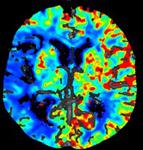
A typical CT perfusion procedure lasts about half a minute and scans the same tissue many times, each scan at a low dose. These scans both reveal the internal anatomy of the patient and show how levels of a contrast agent, such as iodine injected into the bloodstream, change over time. Changing concentrations of iodine can be used to calculate blood volume and flow in order to detect injuries to blood vessels or tumor responses to treatment.
The new adaptive algorithm compares these 20 to 30 scans and can differentiate between anatomical regions that do not change from moment to moment and those regions that carry the contrast agent – effectively reducing image noise while preserving iodine signal. The quality of each scan improves through non-linear comparisons with scans acquired earlier and later in the exam.
At the AAPM meeting, the researchers will present animal data showing the effectiveness of the technique. They have also begun to process data from clinical brain perfusion CT exams in patients.
“We’re up to 15 or 20 cases that we've shown to the docs, and they're all giving us the thumbs up,” McCollough said.
For more information: www.aapm.org

 February 02, 2026