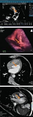
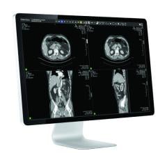
These are the multimodality images (i.e., 2-D, 3-D ultrasound, CT and MR) of a papillary fibroelastoma that were presented online from Memorial Hospital of Tampa, Fla., to a physician in Greenville, N.C., for evaluation for robotic surgery.
In the aviation industry, changing from mechanical flight control systems to fly-by-wire electronic systems improved the capability and safety of airplanes. Like aviation, medicine has witnessed similar technological breakthroughs. In the field of cardiology alone, we’ve seen imaging technology take us from the X-rays and electrocardiograms (ECGs) of yesterday to today’s 2-, 3-, and 4-dimension ECGs, CTs, MRIs and PET/CT fusion scans. Much as pilots can command every component of an airplane from their cockpit seat, I am able to diagnose my patient’s disease from a single workstation.
As a cardiovascular imaging specialist at IASIS Healthcare’s Memorial Hospital of Tampa, Fla., I saw the tremendous value of the advanced visualization tools used on multimodality workstations in radiology and I wanted to bring these tools to my cardiovascular department. IASIS has had great success with McKesson’s Horizon Medical Imaging radiology picture archiving and communications system (PACS) solution, so we turned to McKesson again.
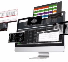
The result is the implementation of Horizon Cardiology, a cardiovascular information solution (CVIS) from McKesson, combined with Vital Images’ Vitrea Enterprise Suite and TomTec’s 4-D Cardio-View. This best-of-breed integrated solution is what I call the “cardiology cockpit,” putting all the controls a cardiologist needs for diagnosis in one location. From this cockpit, I can see the complete cardiac history of a patient. All multimodality images — including multidimensional reconstructions, waveforms and reports — are on a single workstation, enhancing my ability to access, review, compare, manipulate and share them, whether I’m at my hospital or 2,300 miles away at another IASIS facility. Collaboration, confidence and clinical care have been improved.
Pre-Flight
Traditionally, cardiac images have been stored in separate systems and this nonintegrated environment has severely limited our level of diagnostic ability. Cardiologists must travel from one department’s dedicated workstation to the next and the next to view images, making it difficult to comprehend and share the patient’s entire picture. This situation would be like asking an airline pilot to visit the control tower to receive weather and take-off parameters, to visit flight command for weight and fuel details, and to visit maintenance for clearance to fly. Crazy, right? But that’s what we have been doing in the cardiac department.
For instance, I could view and manipulate coronary computed tomography (CT) images on one workstation and conclude my study, but I couldn’t then share the study. Anyone I wanted to collaborate with — cardiac surgeons, fellow cardiologists, referring physicians, medical students —would have to come to that specific workstation or wait to see the saved images on a compact disc (CD) that I gave to them. Our workflow has had cardiologists isolated, reviewing images, dictating reports and sending CDs instead of collaborating. Furthermore, there was no ability to compare those CT images to other studies, such as an echocardiogram or magnetic resonance imaging (MRI). Each patient has a story and cardiologists couldn’t read it completely before the cardiology cockpit became available.
Take-Off
Like many healthcare organizations, IASIS Healthcare has been in a system-wide transition to an electronic health record (EHR) that started in 2004. The goal of this EHR is to replace the paper chart and to centrally store patient data, including images, measurements, waveforms and reports.
As the director of cardiology for the 16-hospital system, I was responsible for selecting a system for the cardiac record. I chose McKesson’s Horizon Cardiology solution — the only single database solution for ECGs, cath, hemodynamic monitoring, echovascular ultrasound, CT, MRI and nuclear cardiology — because its design brings together all the cardiac data onto one workstation to provide the complete picture. For advanced visualization techniques, McKesson provides the vendor-neutral, best-of-class products from Vital Images for manipulating 2-D and 3-D coronary CT images, TomTec for 2-D/3-D/4-D echovascular images and other vendors for nuclear cardiology.
Our cardiology workflow has been transformed because I no longer need to travel from one workstation to another. By combining imaging modalities onto one workstation, I can simultaneously view multiple modalities to see the whole picture of what is going on with the patient. And because the solution offers Web-enabled image and report editing, the patient’s record can be accessed at any time and from any location, whether I’m at the office or at home. Not only can multiple modalities be compared at the same time from the cockpit, but the advanced visualization tools allow for specialized post-processing to highlight and analyze those images.
In-Flight
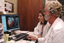
Our cardiology cockpit provides the platform for improved patient care. Recently, a patient with mitral valve problems sat next to me as we called a robotic surgeon from Greenville, N.C. The surgeon was able to remotely view the workstation and look at the transesophageal echo (TEE) to pinpoint that the mitral valve was prolapsing and that it was flail, thereby seeing the leakage for himself. He was reassured that the patient did not need a cardiac cath prior to surgery, since I had done a coronary CT to determine there was no coronary disease. The surgeon commented to the patient the repair would be quite easy. The surgeon was confident, the patient was confident and I was confident. The ability to create that relationship and share the knowledge changes everything in cardiology. Never before could I go to one place to access all of the images and reports and remotely share the information. Now I can seamlessly share images and communicate online with colleagues, and everyone is gaining confidence.
Collaboration also changes the course for the patient. Just because a physician has a report, it may not fully articulate the nuances of the disease, its anatomy and physiology. In the case of our mitral valve patient, she will have better surgical results because of the collaboration. She hand-picked her surgeon, the surgeon was fully informed prior to surgery, and in this case, an unnecessary invasive procedure (the cardiac cath) was avoided.
I was able to determine the information noninvasively, which is economically sound and, most importantly, less risk to the patient. Patients and insurance companies are more at ease and the cardiologist and surgeon are more at ease, because they have the clinical information required for better and safer decisions in patient care.
Nothing But Blue Skies
The cardiology cockpit has tremendous potential for our field, because communication is indispensable. Collaboration with colleagues not only provides a comprehensive understanding of the patient, but also leads to improved clinical outcomes. Today the need for cardiologists to move from workstation to workstation prohibits instantaneous access and comparison of these multimodality images. However, in the future, routine use of 3-D/4-D visualization software to manipulate these images and real-time collaboration with patients and their medical teams will provide better care and less risk. It will reduce reliance on invasive procedures to determine a definitive diagnosis and treatment plan.
At IASIS, the satisfaction of our patients, referring physicians, cardiac surgeons and research team proves daily the value of the Horizon Cardiology cockpit. At IASIS, every day, we cruise cardiac images with our remote imaging partners.
This case study was supplied by McKesson, Vital Images and TomTec.

 March 06, 2024