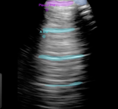
April 24, 2008 - Using 3D ultrasound technology they designed, Duke University bioengineers can compensate for the thickness and unevenness of the skull to see in real time the arteries within the brain that most often clog up and cause strokes.
The researchers believe that these advances will ultimately improve the treatment of stroke patients, whether by giving emergency medical technicians (EMT) the ability to quickly scan the heads of potential stroke victims while in the ambulance or allowing physicians to easily monitor in real time the patients' response to therapy at the bedside.
The results of the latest studies were reported online in the journal Ultrasound in Medicine & Biology. The research was supported by the National Institutes of Health and the Duke Translational Medicine Institute, with assistance from the Duke Echocardiography Laboratory.
"To our knowledge, this is the first time that real time 3D ultrasound provided clear images of the major arteries within the brain," said Nikolas Ivancevich, graduate student in Duke's Pratt School of Engineering and first author of the paper. "Also for the first time, we have been able overcome the most challenging aspect of using ultrasound to scan the brain - the skull."
The key to obtaining these images lies in the design of the transducer. In traditional 2D ultrasound, the sound is emitted by a row of sensors. In the new design, the sensors are arranged in a checkerboard fashion, allowing compensation for the skull's thickness over a whole area, instead of a single line.
"The speed of the sound waves is faster in bone than it is in soft tissue, so we took measurements to better understand how the bone alters the movement of sound waves," Ivancevich explained. "With this knowledge, we were able to program the computer to correct for the skull's interference, resulting in even clearer images of the arteries."
Duke University, biomedical engineering professor Stephen Smith, senior author of the paper, noted "This is an important step forward for scanning the vessels of the brain through the skull, and we believe that there are now no major technological barriers to ultimately using 3D ultrasound to quickly diagnose stroke patients."
"I think it's safe to say that within five to 10 years, the technology will be miniaturized to the point where EMTs in an ambulance can scan the brain of a stroke patient and transmit the results ahead to the hospital," Smith continued. "Speed is important because the only approved medical treatment for stroke must be given within three hours of the first symptoms."
For their experiments, the Duke team studied 17 healthy people. After injecting them with a contrast dye to enhance the images, the researchers aimed ultrasound transducers into the brain from three vantage points - the temples on each side of the head and upwards from the base of the neck. The temple locations were chosen because the skull is thinnest at these points.
Ivancevich took this approach one step further to compensate for the thickness and unevenness of the skull in one subject.
The 3D ultrasound has the benefit of being less expensive and faster than the traditional methods of assessing blood flow in the brain - MRI or CT scanning, Ivancevich said. Though 3D ultrasound will not totally displace MRI or CT scans, he said that the new technology would give physicians more flexibility in treating their patients.
Other Duke members of the team were Gianmarco Pinton of Pratt, and Duke University Medical Center division of neurology researchers Heather Nicoletto, Ellen Bennett and Daniel Laskowitz.
For more information: www.dukenews.duke.edu


 January 28, 2026

