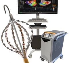
January 21, 2013 — At the Boston Atrial Fibrillation (AF) Symposium 2013, Philips Healthcare introduced its latest innovations in advanced imaging integration for electrophysiologists, showcasing its new EP navigator offering — a novel approach to 3-D rotational angiography that provides real-time anatomical details of cardiac left atrium-pulmonary vein (LA-PV) structures with significantly enhanced workflow.
Furthermore, under the terms of a new agreement with Biosense Webster, Philips plans to offer integration of Philips Allura X-ray images with the CartoAlara Module of Biosense Webster’s Carto 3 electroanatomical mapping system, providing enhanced anatomical detail and orientation for electrophysiologists performing catheter ablation procedures.
Advanced real-time 3-D imaging
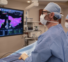
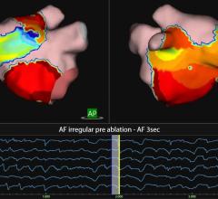
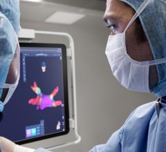
For hospital teams working in catheter ablation, capturing the best anatomical detail possible at time of procedure is essential to accurately guide catheters through the anatomy of a beating heart. Philips’ EP navigator system provides a novel approach to 3-D rotational angiography to capture these images, delivering an alternative to pre-procedural computed tomography/magnetic resonance (CT/MR) imaging to obtain the LA-PV anatomy.
Obtaining 3-D rotational images can be challenging when treating large patients or performing procedures under general anesthesia. Philips’ new EP navigator supports an optimized 3-D rotational scan that significantly enhances the workflow for these procedures, by removing barriers encountered in the traditional workflow. In addition, the optimized 3-D rotational scan requires less radiation exposure to capture the 3-D image, as it uses a shortened trajectory of 159 degrees versus 240 degrees for the traditional scan.
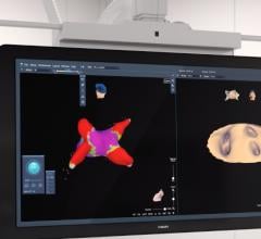
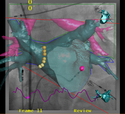
“By using the EP navigator with 3-D rotational scan, my patients and team gain several benefits,” says Vivek Reddy, Boston AF course co-director and director, cardiac arrhythmia service, The Mount Sinai Medical Center. “Firstly, rotational angiography is getting better and better, to the point where we don’t have any issues in getting good quality pictures, so it allows us to optimize workflow. I can now take a rotational image, make a 3-D rendering of it and project it on to the X-ray, so that as we manipulate the catheters on X-ray, the shadows tell us where the catheter is relative to the left atrium.”
Integration with mapping
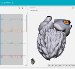
Maximizing the benefits of advanced X-ray imaging and mapping solutions, integration is critical. Philips and Biosense Webster are collaborating to make such integration a reality. Philips EP navigator already supports seamless integration of the LA-PV anatomy obtained through 3-D rotational scans, into Biosense Webster’s Carto 3 mapping system.
Under the terms of the new agreement between Philips and Biosense Webster, Philips plans to offer integration of Philips Allura X-ray images with the CartoAlara module of Biosense Webster’s Carto3 alectroanatomical mapping system. By unifying registered Philips X-ray images into Carto 3 system maps, enhanced anatomical detail and orientation are achieved within a single view.
“At Philips, we are dedicated to make a difference in the treatment of cardiac arrhythmias to improve patient outcomes and quality of life. We continue to advance the tools available to EPs, by offering innovative live image guidance solutions and by collaborating with our industry partners,” said Ronald Tabaksblat, general manager Interventional X-ray at Philips Healthcare. “Our agreement with Biosense Webster perfectly exemplifies this dedication, by allowing Philips Allura X-ray systems to seamlessly integrate with the Carto 3 mapping system.”
The 18th annual Boston AF Symposium took place Jan. 17-19, 2013 at The Seaport Hotel and World Trade Centre in Boston.
For more information:www.philips.com

 September 05, 2025