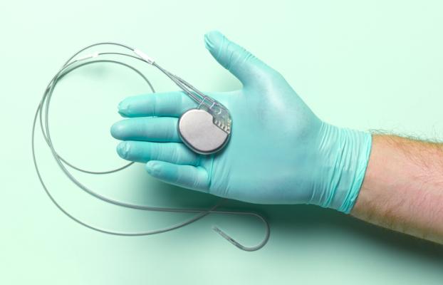
A research study conducted by Children’s Hospital Colorado in Aurora, Colo. found that three-dimensional (3D) echocardiography resulted in more accurate pacemaker lead placement when compared to X-ray technology. The study was presented during the American Society of Echocardiography’s (ASE) 34th Annual Scientific Sessions, held June 23-26 in National Harbor, MD. Image courtesy: Getty Images
July 5, 2023 — Research presented during the American Society of Echocardiography’s 34th Annual Scientific Sessions, ASE 2023, June 23-26, 2023, in National Harbor, Md., shared how advances in echocardiography resulted in more accurately placed pacemaker devices.
The study, titled 3D Echocardiography Guidance for Pacemaker Lead Placement Improves Accuracy of Lead Placement and Reduces QRS Duration Compared to Fluoroscopic Guidance, was presented June 25 during ASE 2023, and one of nearly 400 abstract poster presentations featured during the event to highlight continuing innovations in the field of cardiovascular ultrasound.
Some pacemakers use flexible, insulated wires, or leads, to deliver electrical pulses that help hearts beat at a normal rate and rhythm. As summarized in an online ASE 2023 news summary, the current standard practice for placing pacemaker leads is fluoroscopic/X-ray guidance. However, a new research study conducted by Children’s Hospital Colorado in Aurora, Colo., found that three-dimensional (3D) echocardiography resulted in more accurate pacemaker lead placement when compared to X-ray technology.
The study’s lead author Dale Burkett, MD, Assistant Professor in Pediatrics-Cardiology at Children’s Hospital Colorado, explained that when pacemaker leads are placed in the ideal location, they are able to better mimic the heart’s normal electrical conduction and reduce the long-term risk of heart dysfunction. Additionally, noted the ASE’s research spotlight feature, 3D echocardiography lowers both the patient’s and the cardiology staff’s exposure to radiation during the procedure.
“By using 3D echocardiography guidance, we are able to better visualize pacemaker leads as they move through the heart and guide them to where they are intended to go,” said Burkett, adding, “Our work demonstrates that with advances in echocardiography, we can provide higher quality care for our patients who need permanent pacemakers for heart rhythm management.”
A summary of the methodology, results and conclusion presented follow:
Detailing the methodology, the team noted that in 59 patients, 3DE was used to guide pacemaker leads to desired locations, where they were secured; fluoroscopy was used sparingly as needed for lead movement, securing leads, or slack adjustment. Lead location was recorded by echocardiography, and (in those that had it) CT or MRI. Paced QRS duration was measured in RV leads. Cases were compared to 72 historical control pacemaker cases, which used only fluoroscopy for lead implant, by Wilcoxon rank sum, Pearson’s chi-square or Fisher’s exact tests.
In writing of their results, the authors offered this: When RV lead location was assessed by echocardiography, using 3DE guidance, 78.4% of RV leads were placed in the septum, 17.7% were placed in the junction between septum & free-wall, and only 3.9% were placed in the free-wall, compared to 29.8%, 12.8% and 57.4% by fluoroscopic guidance, respectively.
They added that this trend was also seen in the limited number of patients that had a subsequent CT or MRI. Of the two RV leads placed in the free-wall while using 3DE guidance, one was an ICD-only lead, and in the other, the ICD+pacemaker lead was guided to the septum but pacing parameters were not tolerable and thus the free-wall was used. QRS duration was significantly shorter in patients that had 3DE guidance with the lead placed in the septum (129msec, IQR 117-143) compared to those with fluoroscopic guidance alone (141.5msec, IQR 134-148) (P=0.031).
The researchers concluded that the use of 3DE guidance for pacemaker lead placement results in more accurate placement in the septum and shorter QRS duration than fluoroscopic guidance, adding that 3DE guidance for lead placement could reduce the long-term risk of pacemaker-induced cardiomyopathy and should be considered for all pacemaker lead placements.
The study was presented as part of the 2023 Arthur E. Weyman Young Investigator’s Award Competition, where it was a finalist. Additional researchers conducting the study with Burkett included: Kathryn Collins, MD, Dustin Nash, MD, Martin Runciman, MBBS, Johannes von Alvensleben, MD, all of Children's Hospital Colorado, (Aurora, CO); and Pei-Ni Jone, MD, of Lurie Children's Hospital of Chicago (Chicago, IL).
Follow DAIC’s coverage of ASE 2023 news here.
More information: www.ASEcho.org, or asescientificsessions.org

 June 08, 2023