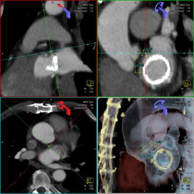
Figure 3: syngo DynaCT Cardiac with syngo iGuide Toolbox markers indicating the positions of the paravalvular leaks from TEE.
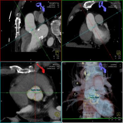
Figure 4: syngo DynaCT Cardiac with syngo iGuide Toolbox markers indicating the plane of the aortic valve.
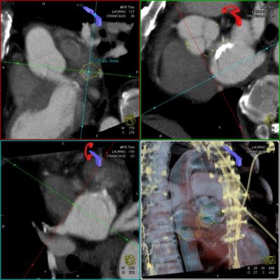
Figure 5: syngo DynaCT Cardiac with syngo iGuide Toolbox markers indicating the desired region of the transseptal puncture.
Patient History
72-year-old, male patient with previously placed bioprosthetic mitral valve.
Diagnosis
Mitral regurgitation (grade 3+) due to periprosthetic leak (Video 1). To view the videos click here.
Treatment
Closure of the paravalvular leak with an 8 mm VSD closure device under TEE (transesophageal echo) and fluoroscopic guidance. syngo iGuide Toolbox graphics were overlaid as additional guidance.
Comments
The case demonstrated that the overlay of 3-D information on the live fluoroscopic images provided additional helpful information.?? More detailed information can be found in the following publication:? Krishnaswamy A, Tuzcu EM, Kapadia S. "Three-dimensional computed tomography in the cardiac catheterization laboratory." Catheter Cardiovasc Interv. 2010
Detailed Workflow Description
Pre-procedural cardiac CT data was retrieved from the PACS. (Video 2) The following structures were marked with syngo iGuide Toolbox graphics:
• Mitral valve plane
• Positions of the paravalvular leaks (based on the information of the diagnostic TEE Picture 3 )
• Aortic valve plane (Picture 4)
• Area for transseptal puncture (Picture 5)
?
The patient was positioned on the table with arms up to match the patient’s position during the pre-procedural cardiac CT. Arms were kept up during the whole procedure. A five-second syngo DynaCT Cardiac run was performed without contrast (Video 6). During this rotational acquisition a pigtail catheter was positioned in the aortic root to visualize the position of the aorta and the aortic root as landmarks for the syngo InSpace 3-D/3-D registration. Patient was asked just to hold his breath, i.e., no deep inspiration or expiration in order to be in a similar breathing situation as during the remaining procedure. This helps increasing the overlay accuracy.?? 3-D/3-D registration of syngo DynaCT Cardiac image and CT was performed in syngo InSpace with the pigtail catheter and the mitral valve serving as landmarks. (Video 7).??
The overlaid 3-D markers helped as additional guidance during the procedure for:
• a) the puncture of the atrial septum
• b) the progression of a guidewire through a leak (Video 8)
• c) the adjustment of the closure devices (Video 9)

 December 24, 2025


