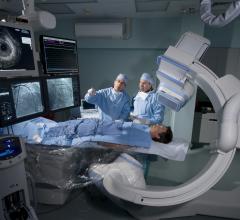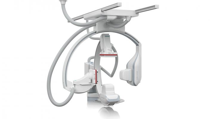
November 26, 2012 — At the 98th annual meeting of the Radiological Society of North America (RSNA) in Chicago, Siemens Healthcare is displaying for the first time its new X-ray tube and detector technology for the Artis Q and Artis Q.zen angiography systems, designed to improve minimally invasive therapy of diseases such as coronary artery disease, stroke and cancer.
The new X-ray tube in both the Artis Q and Artis Q.zen series is intended to help physicians identify small vessels up to 70 percent better than conventional X-ray tube technology. The Artis Q.zen combines this innovative X-ray source with a new detector technology designed to support interventional imaging in ultra-low-dose ranges to patients, doctors and medical staff, particularly during longer interventions.
The second generation of Siemens’ flat emitter technology is key to the advances made in the X-ray tube for the Artis Q and Artis Q.zen product lines. Instead of the coiled filaments used in conventional X-ray tubes, flat emitter technology is used exclusively in the new tube to emit electrons. Flat emitters are designed to enable smaller quadratic focal spots that lead to improved visibility of small vessels by up to 70 percent. Both physicians and patients benefit from a high level of detail in imaging-supported interventional therapy. Neurologists can more precisely measure blood circulation in specific areas of the brain, for example, while stenoses in the heart’s smallest blood vessels can be spotted in coronary angiography.
Examinations using ultra-low-dose radiation
The Artis Q.zen series has been designed to combine the X-ray tube with a detector technology that allows detection at ultra-low radiation levels. Artis Q.zen imaging can use doses as low as half the standard levels applied in angiography. This improvement is the result of several innovations, including a fundamental change in detector technology. Until now, almost all detectors have been based on amorphous silicon. The new crystalline silicon structure of the Artis Q.zen detector is designed to be more homogenous, allowing for more effective amplification of the signal, greatly reducing the electronic noise even at ultra-low doses.
The Artis Q.zen was developed to support better imaging quality at ultra-low-dose ranges, reducing the radiation exposure of patients, physicians and medical staff. This is especially important in dose-sensitive application fields such as pediatric cardiology and radiology, or electrophysiology, which is being used more frequently as rates of cardiac arrhythmia increase in an aging population.
Innovative applications for interventional imaging
In addition to the hardware, several software applications have been designed to improve interventional imaging. In coronary artery disease treatment, the applications allow precise correlation of angiography images with ultrasound images taken by a probe inside the coronary arteries. Stents are imaged in real time during therapy, with motion stabilization created by simultaneous correction for the heartbeat.
Other new 3-D applications are designed to image the smallest structures inside the head. Their high spatial resolution is crucial for imaging intracranial stents or other miniscule structures such as the cochlea in the inner ear. Moving organs such as the lungs can be imaged in 3-D in less than three seconds, reducing motion artifacts and the required amount of contrast agent. Through visualization and measurement of blood volumes in the liver or other organs, Siemens’ functional 3-D imaging provides a basis for planning therapies such as chemo-embolization of hepatic tumors.
The Artis Q and Artis Q.zen are pending U.S. Food and Drug Administration (FDA) 510(k) clearance and are not yet commercially available in the United States.
For more information: www.siemens.com/healthcare

 October 24, 2025