October 30, 2018 — At 18 weeks, a baby’s heart is the size of an olive and beating about 150 times per minute.[1] The structure itself is extremely complex — and with the baby in constant motion, it is always a moving target.
Evaluating the fetal heart is complicated. In fact, the initial assessment can be one of the most difficult ultrasound exams to perform, particularly for less-experienced users with minimal training.
However, these assessments are critical to detect complications or anomalies, such as congenital heart defects which affect one out of every 110 babies born around the world[2]. To complicate matters, most women are completely blindsided by the diagnosis, as 90 percent occur in pregnancies where there are no known risk factors.[3]
About three years ago, Greggory DeVore, M.D., a specialist in maternal fetal medicine at Huntington Hospital, Pasadena, Calif., became particularly interested in fetal heart assessment when he came across a software program that was used by adult and pediatric cardiologists to examine the heart’s function using speckle tracking analysis – a technique that analyzes the motion of tissues in the heart.
He thought to himself, “I wonder if I could do that in the fetus.”
At the time, the only tools available were used to look at the anatomy and heart rate, but he wanted to also know how the heart’s shape, size and how it was contracting.
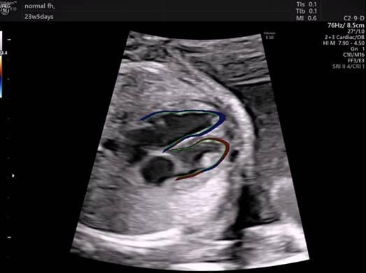
DeVore acquired the software, and that same day, he began looking at fetal hearts. He used the measurement tools for the adult heart but found that they were not helpful to the fetal evaluation. With some help, DeVore reprogrammed the software to divide the fetal heart in 24 segments – measuring the heart in ways that had never been done before.
“This was the genesis of the creativity behind using this software,” DeVore said. “From this, we made several measurements of the heart’s size, shape and contractility – or how it’s squeezing. We immediately got to work and published 13 peer-reviewed articles that described the clinical value of this software.”
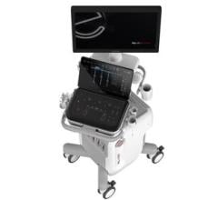
This new tool – called fetalHQ – runs on GE Healthcare’s Voluson ultrasound systems and is the first tool to simultaneously examine the size, shape and function of the fetal heart.
Watch a VIDEO example of this technology
“This is an incredible step forward in examining the fetal heart. Previously, we had various tools that looked at specific sites of the heart – but nothing that examined the entire chamber,” DeVore said. “On top of this, the heart had to be in a specific position – at 12 or six o’clock on the screen to get the proper measurements. And this all required an advanced level of diagnostic skills to learn the techniques.”
Congenital heart defects are also among the most difficult fetal abnormalities to detect. In general, low-risk detection rates are between 30 to 50 percent.[4]
For example, co-arctation of the aorta is a narrowing of the aorta, the large blood vessel that branches off the heart and delivers oxygen-rich blood to the body. When this occurs, the heart must pump harder to force blood through the narrowed part of the aorta.
When examining the fetal heart’s anatomy, there are findings that are associated with coarctation, but are not always 100 percent specific for this condition. Because of this, the clinician often has to play a guessing game to decide whether coarctation is present or not. By looking at the function rather than the structure with fetalHQ, DeVore found he was able to separate these cases with almost 100 percent accuracy.
In addition, DeVore described cases where the fetus was not growing properly. Typically, this is defined as a weight less than the tenth percentile, and then divided into three groups. One of these groups entails fetuses that are between the third and tenth percentile with normal blood flow to the placenta and brain. While small, these pregnancies are typically deemed “normal” with no risk factors.
However, when DeVore examined 50 of these cases with fetalHQ, he discovered abnormal cardiac function. “This changes the whole paradigm as to how you interpret and manage these types of fetuses going forward,” DeVore said. “It allows us to ask, ‘How is the heart working?’ We’re now able to identify abnormal function, shape and size of the heart – things we couldn’t see previously.”
“Perhaps, the most incredible feature of this new tool is that we are able to collect all this information – the size, shape, and function of the fetal heart – in just two minutes and thirty seconds,” DeVore concluded.
For more information: www.gehealthcare.com
References:
1. https://www.parents.com/pregnancy/week-by-week/18/your-growing-baby-week-18/
3. Why screen for heart defects?
4. Cost-effectiveness of prenatal screening strategies for congenital heart disease

 January 28, 2026
