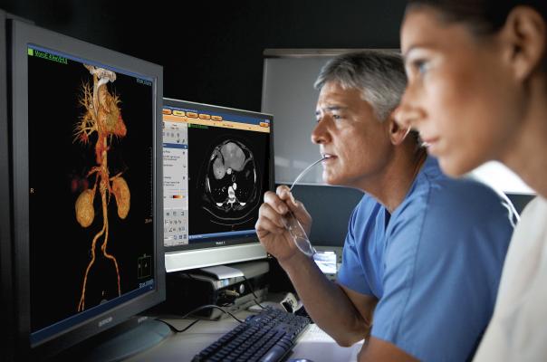
Computed tomography has seen rapid growth in all segments of medicine over the past two decades, and recent advances may further expand use of cardiac computed tomography angiography (CCTA). Advocates for cardiac CT speaking at the 2015 meetings of the American College of Cardiology (ACC), Society of Cardiovascular Computed Tomography (SCCT) and Transcatheter Cardiovascular Therapeutics (TCT) laid out the latest technological advancements and outlined new clinical trial data that may help CT become a first-line, one-stop-shop cardiac imaging modality in the near future.
Advances in CCTA include perfusion imaging, spectral imaging, noninvasive fractional flow reserve CT (FFR-CT) and new technologies that have greatly lowered cardiac CT radiation dose levels, said James Min, M.D., professor of radiology and medicine and director of the Dalio Institute of Cardiovascular Medicine, Weill Cornell, New York Presbyterian Hospital.
Min said the goal of any diagnostic system that evaluates coronary disease should be anatomic assessment, the ability to offer an accurate prognosis, identify specific culprit lesions causing ischemia and aid in plans for revascularization. Today, he said CTA offers all of these tools in one test and may be more cost-effective than the current standard of care involving numerous diagnostic exams, overnight stays and diagnostic catheter angiographic assessments.
VIDEO - Implementation and the Science Behind FFR-CT
VIDEO - The Status FFR-CT Adoption in the United States
Noninvasive CT-FFR Assessments
In November 2014, the U.S. Food and Drug Administration (FDA) FFR-CT software to noninvasively, virtually assess the blood flow through plaque narrowings of coronary vessels to see if the lesions have any hemodynamic significance. This may eliminate the need to perform invasive FFR readings in the cath lab, since the FFR-CT has a close correlation with the catheter-based technology. Catheter FFR is currently the gold standard for assessing the functional significance of lesions and to justify the need for stents. Many luminaries at SCCT and TCT see FFR-CT as a potential paradigm shift in how chest pain and cardiac patients will be assessed in the future. This technology was the primary headliner for sessions at SCCT 2015, and the subject of numerous packed sessions.
A handful of U.S. centers are now implementing FFR-CT, and several studies are underway to further validate its comparison with invasive FFR and its cost-effectiveness.
“Up to half of all diagnostic heart catheterizations in the United States reveal no significant coronary artery disease,” said Gil Raff, M.D., director of cardiac MRI and CT at Beaumont Hospital — Royal Oak, Mich., one of the centers to adopt FFR-CT. “FFR-CT gives us the individual patient information we need without having to perform an invasive procedure. This means we are able to reduce costs, along with the risk of radiation exposure and possible patient complications.”
HeartFlow’s FFR-CT is a Web-based, computation fluid dynamics software that analyzes existing CCTA images to provide physicians with detailed data about blood flow within the coronary arteries. It uses a standard noninvasive coronary CT angiography scan, uploaded to the cloud, and combines it with proprietary computer algorithms to create a three-dimensional model of the patient’s coronary arteries that can determine whether any given plaque requires cardiac catheterization.
Results from the PLATFORM study showed FFR-CT may reduce costs in selected symptomatic patients with suspected coronary artery disease. The cost and quality of life outcomes data were reported at the 2015 TCT meeting. In the 584 patients, 74 percent had atypical angina, and the pre-test probability of coronary disease was 49 percent. In the planned invasive stratum, mean costs were 32 percent lower among the FFR-CT patients than among the usual care patients who typically underwent invasive coronary angiography ($7,343 vs. $10,734, p<0.0001). In the noninvasive stratum, mean costs were not significantly different between the FFR-CT patients and the usual care patients who underwent traditional noninvasive risk stratification ($2,679 vs. $2,137).
In a sensitivity analysis, when the cost weight of FFR-CT was set to seven times that of CTA, the FFR-CT group still had lower costs than in the usual care group in the invasive testing stratum ($8,619 vs. $10,734). In the noninvasive testing stratum, when the cost weight of FFR-CT was set to half that of CTA, the FFR-CT group had higher costs than the usual care group ($2,766
vs. $2,137).
“These results suggest that use of FFR-CT might reduce overall costs and improve patient quality of life,” said lead researcher Mark Andrew Hlatky, M.D. He is professor of health research and policy and of medicine at the Stanford University School of Medicine in California. “Larger, randomized studies are warranted to compare the clinical effectiveness of management strategies based on use of FFR-CT with management strategies based on using other methods of anatomic or functional evaluation.”
Spectral CT Imaging
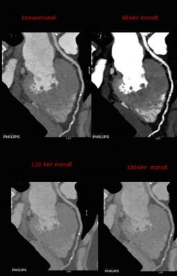
There has been a renewed interest in the development of spectral CT over the past couple years. Spectral imaging allows viewing of the same anatomy at two different kV energies. Different anatomical features are enhanced and easier to see at different energy levels. Additionally, spectral software can highlight or eliminate specific chemical compounds based on their atomic number, including iodine and calcium. This allows differentiation between calcified coronary lesions and iodine contrast in the blood vessel, or enables reconstruction of contrast and non-contrast images from a single contrast scan.
In the past, this meant using a dual-source CT scanner to image a patient at two different energy levels. Today, Philips, GE and Siemens have introduced new, single-source CT scanners that enable dual-energy imaging in a single scan. This is accomplished though dual-layer detectors with elements that absorb different energy levels and fast kV switching on the X-ray tube during scanning. The newest version of Siemens’ Somatom Definition Edge CT acquires dual-energy images using a single-source X-ray tube that emits two energy spectra simultaneously.
In September, the U.S. Food and Drug Administration (FDA) granted 510(k) for Philips’ Spectral Diagnostic Suite (SpDS), which offers a set of advanced visualization and analysis tools designed for the IQon Spectral CT technology. It allows clinicians to utilize the spectral information on-demand, without the added complexity of special modes or workstations that disrupt user workflow. Additionally, because there is no need to bring the patient back for additional imaging, on-demand spectral analysis of a particular region allows the physician to further analyze incidental findings. The package includes spectral enhanced comprehensive cardiac analysis and spectral enhanced advanced vessel analysis.
The Rise of CT Perfusion Imaging
There is mounting clinical evidence from numerous trials showing CT perfusion imaging can accurately assess functional blood flow in the heart without the need for a nuclear exam, stress testing or magnetic resonance imaging (MRI). CT system vendors and third party advanced visualization vendors TeraRecon and Vital Images have all released perfusion assessment software, which present various maps of iodine contrast in the blood as it flows through the myocardium. The technology may reduce or eliminate the need for nuclear myocardial perfusion exams, which are currently the gold standard for perfusion assessment.
“Stress myocardial CT perfusion imaging has been rapidly validated and I think we have come a long way in just five years of trials,” said SCCT President Ricardo Cury, M.D., FAHA, FSCCT, FACC, chairman of radiology, director of cardiac imaging, Baptist Health of South Florida, Miami.
Related content on CT Perfusion imaging:
Watch a VIDEO on CT perfusion advances from SCCT 2015
Advances in CT Perfusion Imaging
New Technology Supports CT as Prime Cardiac Imaging Modality
PROMISE Trial Shows CT is as Effective as Stress Tests and SPECT to Assess Chest Pain
Developing CAD-RADS Measures For Cardiac CT
The American College of Radiology (ACR) has developed several standardized reporting and data system (RADS) measurements of various disease states and trauma assessments, including Lung-RADS for lung cancer, BI-RADS for breast assessments and HI-RADS for head injuries. SCCT is working with ACC and ACR to develop a CAD-RADS risk score system for coronary artery disease assessments via CCTA. Cury said this will be a major effort for CCTA to bring it in line with the movement toward valve based imaging. The system will have scoring levels 1-5 and each level reported will link to a set a patient management recommendations.
VIDEO - The Introduction of CAD-RADS
 May 12, 2020


