May 26, 2022 — At the 69th Annual Conference of the Israel Heart Society, UltraSight, an Israeli-based digital health ...
Cardiovascular Ultrasound
This channel includes news and new technology innovations about cardiovascular ultrasounds. Cardiovascular ultrasounds, or echocardiograms, use ultrasound imaging to provide a picture of the heart.
 May 26, 2022
May 10, 2022 — MedTech Breakthrough, an independent market intelligence organization that recognizes the top companies ...
May 10, 2022
The global cardiovascular ultrasound systems market was expected to register a CAGR of 5.9% over the forecast period ...
May 05, 2022Sponsored Content

Blog | Artificial Intelligence
New research shows superiority of Ultromics’ AI in predicting cardiac-related death As the use of artificial ...
October 10, 2022
April 29, 2022 – Aurora St. Luke’s Medical Center in Milwaukee is the first site in the country to join a clinical trial ...
April 29, 2022
April 21, 2022 – Coronary artery disease (CAD) is one of the major causes of mortality and morbidity worldwide ...
April 21, 2022
April 20, 2022 – Researchers from the RIKEN Center for Advanced Intelligence Project (AIP) and colleagues have tested ...
April 20, 2022Sponsored Content

Case Study | Cardiovascular Information Systems (CVIS)
Washington Health System (WHS) provides healthcare services at more than 40 offsite locations across three counties in ...
April 01, 2021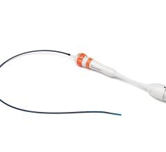
April 12, 2022 – Siemens Healthineers, the global leader in intracardiac echocardiography (ICE), announced the market ...
April 12, 2022![Royal Philips, a global leader in health technology, today announced the launch of Ultrasound Workspace [OLK1] at the American College of Cardiology’s Annual Scientific Session & Expo (ACC 2022).](/sites/default/files/styles/content_feed_medium/public/philips-ultrasound-workspace-cardiology-diagnosis.jpeg?itok=VFUpaaT2)
April 2, 2022 — Royal Philips, a global leader in health technology, today announced the launch of Ultrasound Workspace ...
April 02, 2022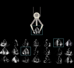
March 31, 2022 – DiA Imaging Analysis Ltd, a leading global provider of AI-powered ultrasound analysis software ...
March 31, 2022Sponsored Content
Case Study | Cardiovascular Ultrasound
As part of the Consolidated Appropriations Act of 2018, pass-through payment status for LUMASON® (sulfur hexafluoride ...
May 29, 2019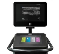
March 9, 2022 — Fujifilm Sonosite, Inc., a world leader in point-of-care ultrasound (POCUS) solutions today announced it ...
March 09, 2022
February 14, 2022 – Clarius Mobile Health has introduced a third-generation product line of high-performance handheld ...
February 14, 2022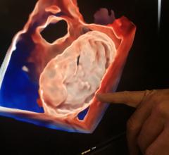
January 17, 2022 – As the increasing number of structural heart interventions are assisted by real-time imaging guidance ...
January 17, 2022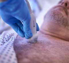
December 9, 2021 – Philips Healthcare expanded its cardiac ultrasound portfolio with new imaging tools and features to ...
December 09, 2021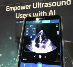
December 2, 2021 — Artificial intelligence (AI) vendor DiA Imaging Analysis was featured in a recent study presented by ...
December 02, 2021
Examples of TrueView and GlassView 3D cardiac ultrasound visualization and artificial intelligence (AI) assisted ...
November 24, 2021