Getty Images
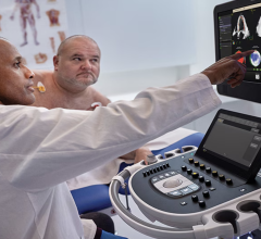
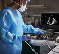
June 29, 2021 – Three research studies presented at the American Society of Echocardiography (ASE) 2021 virtual meeting, June 18-21, highlight the key role of echocardiography in the diverse field of women’s health.
• One study used echo to demonstrate a link between high blood pressure in pregnancy and future microvascular disease.
• Another investigation evaluates the function of the left atrium in women who take trastuzumab, an antibody used in the treatment of breast cancer.
• A third study highlights a heart abnormality seen on routine obstetric ultrasounds associated with significant heart defects, and the value of referring these expectant moms to a center with specialized expertise in fetal cardiovascular conditions.
Echo Shows Link Between Pregnancy Hypertension and Future Microvascular Disease
Women with a history of high blood pressure during pregnancy are more likely to develop subsequent adverse heart function, according to a new study titled Association of Hypertensive Disorders of Pregnancy with Coronary Microvascular Function 8-10 Years after Delivery. Researchers hypothesized that high blood pressure problems during pregnancy might indicate future small vessel disease in the mother’s heart. A special type of echocardiogram using contrast was used to determine that women with a history of high blood pressure disorders during pregnancy do indeed have impaired blood flow in the heart eight to 10 years after delivery.
“Women with a history of hypertensive disorders of pregnancy may be at higher risk of coronary microvascular dysfunction, abnormalities in the small blood vessels in the heart identified by contrast echocardiography, in the decade after their delivery. These findings persist even after adjusting for other cardiovascular risk factors. From prior studies, we know that women with hypertensive disorders of pregnancy are at increased risk for heart disease later in life. Identifying those who have microvascular dysfunction could help guide therapeutic options to modify this risk,” explained lead author Malamo Countouris, M.D., University of Pittsburgh Medical Center Heart and Vascular Institute
Trastuzumab for HER2+ Breast Cancer Associated With Reduced Ejection Fraction
While prior research has shown that trastuzumab therapy for HER2+ breast cancer can be associated with reduced left ventricle ejection fraction (EF), the effect of this therapy on function of the left atrium remains largely unknown. In their novel study Left Atrial Function is Reduced Following Trastuzumab in Breast Cancer Patients, researchers used echocardiography with speckle tracking to evaluate the effects of this known cardiotoxic treatment on 168 women over the course of a year.
“Most work has gone into studying how trastuzumab affects systolic function, and left ventricular ejection fraction is measured before and during treatment. However, left atrial function may possibly be affected before left ventricular systolic function is impaired and thus may serve as an early marker of cardiotoxicity," said lead author Mats H. Lassen, M.D., Gentofte University Hospital, University of Copenhagen. "In our study we were able to show that left atrial function, as assessed by speckle tracking echocardiography, did in fact decline following trastuzumab treatment. These results could possibly be used as a therapeutic guide in patients receiving trastuzumab, as the sensitive nature of left atrial strain could allow for early detection of cardiotoxicity and thus lead to earlier use of cardioprotective drugs. Our findings could lead to a change in how we evaluate therapy in the treatment of breast cancer. The long-term implications, including development of atrial arrhythmias, warrant further investigation.”
Heart Abnormality on Routine Obstetric Ultrasounds Associated With Significant Heart Defects
One of the most common reasons for patient referral to a fetal heart center is finding on a routine obstetrical ultrasound that the right ventricle (RV) of the fetal heart appears disproportionately larger than the left ventricle (LV). While some degree of RV-LV disproportion is typical in utero, an exaggerated ratio can be associated with serious heart defects. Research study Fetal Diagnosis of Right Ventricle-Left Ventricle Size Disproportion – What Comes Next? looked at outcomes for patients flagged with this abnormality in order to determine the significance of this early sign. As it turns out, 44% of these patients ended up needing surgical intervention after birth, and only about 21% were born with functionally normal hearts.
“Our study is important as it helps guide referring obstetricians concerned about the appearance of the fetal heart on routine ultrasound screening as to the various post-natal outcomes and possibilities, some of which are benign and others which require intervention and post-natal care," said lead author Meghan Kiley Metcalf, M.D., Children's Hospital of Philadelphia. "The shape of the left ventricle, in particular its width, is significantly associated with the need for surgery after birth. This discovery can provide guidance to parents and obstetricians, and will help streamline referral for expert fetal cardiovascular evaluation, thus improving utilization of healthcare resources and outcomes.”
These posters will be presented as a part of the ASE 2021 Scientific Sessions online.
For more information: ASEcho.org


 June 12, 2024


