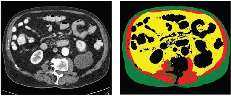
70-year-old White male patient with weight of 79.8 kg, BMI of 29.3, low cardiovascular risk factors (nonsmoker, no diabetes diagnosis, blood pressure of 120/78). Left: Axial CT image at level of L3 vertebral body. Right: Matching automated segmentation label map. Visceral fat area z score is 1.41, corresponding to the 92nd percentile. Patient experienced both subsequent myocardial infarction and stroke.
August 31, 2022 — According to ARRS’ American Journal of Roentgenology (AJR), fully automated and normalized body composition analysis of abdominal CT has promise to augment traditional cardiovascular risk prediction models.
“Visceral fat area from fully automated and normalized analysis of abdominal CT examinations predicts subsequent myocardial infarction or stroke in Black and White patients, independent of traditional weight metrics, and should be considered as an adjunct to BMI in risk models,” wrote first author Kirti Magudia, MD, PhD, currently from the department of radiology at Duke University School of Medicine.
Dr. Magudia and colleagues’ retrospective study numbered 9,752 outpatients (5,519 women, 4,233 men; 890 self-reported Black, 8,862 self-reported White; mean age, 53.2 years) who underwent routine abdominal CT at Brigham and Women’s Hospital or Massachusetts General Hospital from January–December 2012, sans a major cardiovascular or oncologic diagnosis within 3 months of examination. Fully automated deep learning body composition analysis was performed at the L3 vertebral level to determinate three body composition areas: skeletal muscle area, visceral fat area, and subcutaneous fat area. Subsequent myocardial infarction or stroke was established via electronic health records.
Ultimately, after normalization for age, sex, and race, visceral fat area derived from routine CT was associated with risk of myocardial infarction (HR 1.31 [1.03–1.67], p=.04 for overall effect) and stroke (HR 1.46 [1.07–2.00], p=.04 for overall effect) in multivariable models in Black and White patients; normalized weight, BMI, skeletal muscle area, and subcutaneous fat area were not.
Noting that their large study demonstrates a pipeline for body composition analysis and age-, sex-, and race-specific reference values to add prognostic utility to clinical practice, “we anticipate that fully automated body composition analysis using machine learning could be widely adopted to harness latent value from routine imaging studies,” the authors of this AJR article concluded.
For more information: www.arrs.org

 January 28, 2026