Medical imaging plays a key role in cardiology, and most of the newest radiology technology advances are first unveiled at the world’s largest radiology conference, the Radiological Society of North America (RSNA) meeting held each November in Chicago. RSNA 2017 included several key technology advances in several modalities that will impact cardiovascular imaging and will likely be discussed or shown at the key cardiology meetings in 2018.
Watch the following video overview of the new medical imaging technologies shown at RSNA 2017, "VIDEO: Editor’s Choice of the Most Innovative News Technologies."
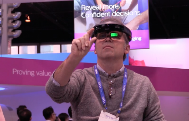
Augmented Reality for Interventional, Surgical Procedures
The most interesting new technology at RSNA related to cardiology were several demonstrations by vendors showing how augmented reality may be able to enhance cath lab and surgical procedures. These systems use a Microsoft HoloLens head visor to project clinical images, including 3-D, in mid-air above a patient, or registered with and projected inside a patient.
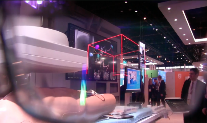
Philips Healthcare had the most impressive display of this work-in-progress technology to improve efficiency and navigation by integrating augmented reality with its angiography interventional guided therapy systems (AR-IGT). The physician can see everything in the room as normal with the visor on, but the HoloLens can project virtual screens to see patient information, hemodynamics information, or to open a virtual control panel for table or C-arm movements without physically touching the tableside controls. Supplemental imaging can be opened in a virtual window in front of the physician without them needing to look at the conventional overhead screen.
Additionally, 3-D reconstruction images of the anatomy from computed tomography (CT) scans or rotational angiography can be projected as virtual holograms anywhere in the room or above the patient. The physician can use voice commands or mid-air hand movements to rotate the 3-D images on any axis, or can move their head or walk around the image projection to see it from different angles. Seeing these images in true 3-D rather than on flat 2-D screens significantly changes perspective and may offer improved understanding and navigation of complex anatomy. Live echo images can also be projected in the same manner and positioned over live fluoro images.
Another feature of the system is it can automatically identify objects picked up and viewed with the HoloLens visor. If the physician picks up an ultrasound transducer it can automatically open an imaging window in the visor to see the live echo images. If a device’s packaging box is held up and viewed, the system can record its use in the procedure log. It also can open a window showing what the device is and any key product information, such as inflation and bursting pressures for angioplasty balloons. It also offers an option to view the device instructions for use.
Philips was showing the prototype system still in development to gather feedback from physicians. While to some clinicians the extra tech may appear cumbersome and unnecessary, the system is intuitive to use and offers the physician complete, hands-free control of their interventional room. It has the ability to pull up imaging or other information without relying on other people. However, the chief benefit will likely be the ability to view true 3-D anatomical imaging for improved procedural navigation while simultaneously being able to see everything else in the room.
Novarad and TeraRecon also showed augmented reality systems in their booths using the HoloLens system that projects either multiplanar image datasets from CT or magnetic resonance imaging (MRI), or 3-D reconstructions of the patient anatomy. By using mid-air hand movements or voice commands, users can scroll through the dataset image slices or rotate and enlarge the 3-D images. The technology allows a surgeon to annotate the patient's skin while viewing structures inside the patient.
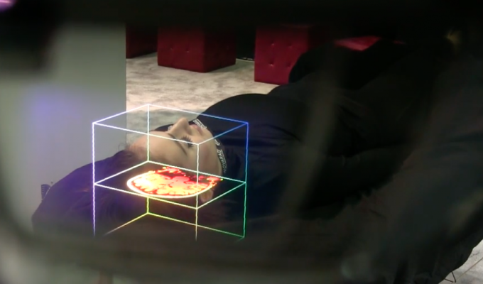
Novarad’s OpenSight augmented reality for surgical applications was demonstrated with a live person laying on a table in their booth to simulate a patient on an operating room or cath lab table. The simulated patient had the medical images registered with their anatomy so the HoloLens user could virtually slice through the patient to examine the anatomy of interest overlaid on the patient. Novarad said the technology may aid procedural planning and allow better visualization of internal anatomical landmarks.
There were usually several attendees waiting to try out each of these systems each time I walked past the Philips and Novarad booths.
Artificial Intelligence Floods Radiology
RSNA 2017 will be remembered as the year artificial intelligence (AI) exploded in radiology. It was by far the biggest news and overall trend in sessions and throughout the expo floor. The key message was AI is here to help and augment radiologists, not replace them. The majority of the AI products demonstrated on the show floor run in the background of radiology IT systems to aid workflow, not offer its own clinical findings. However, several deep learning systems demonstrated had the ability to identify abnormalities in medical images prior to a radiologist reading the study and flag them for priority reads. Auto-diagnostic software was also demonstrated for CT scans for stroke and lung abnormalities.
Here are links to ITN’s coverage of AI at RSNA 2017:
Technology Report: Artificial Intelligence at RSNA 2017
VIDEO: Examples of How Artificial Intelligence Will Improve Medical Imaging
VIDEO: Deep Learning is Key Technology Trend at RSNA 2017
VIDEO: How Utilization of Artificial Intelligence Will Impact Radiology
Why AI By Any Name Is Sweet For Radiology
GE Healthcare and NVIDIA announced a series of imaging equipment advances powered by NVIDIA’s AI computing platform. The announcements included the new Revolution Frontier CT system, advancements to the Vivid E95 4-D ultrasound and development of GE Healthcare’s Applied Intelligence analytics platform. The Vivid E95 4D Ultrasound System uses NVIDIA GPUs to provide fast, accurate visualization and quantification while streamlining workflows across the cSound imaging platform. The GPUs accelerate reconstruction and visualization of blood flow and improve 2-D and 4-D imaging for echo lab and interventional deployments. NVIDIA is working with GE Healthcare to spread its application in healthcare. GPU-accelerated deep learning solutions can be used to design more sophisticated neural networks for healthcare and medical applications — from real-time medical condition assessment to point-of-care interventions to predictive analytics for clinical decision-making. For patients, the partnership aims to drive lower radiation doses, faster exam times and higher quality medical imaging.
Fujifilm Medical Systems announced the expansion of the company's AI development to enhance its informatics healthcare portfolio, which includes Synapse Cardiovascular.
Arterys Inc. unveiled its Arterys MICA platform, an AI platform for medical image analytics. Today, most imaging interpretation solutions are installed inside hospitals and lack the computing power to deliver real-time, interactive AI insights to medical images, according to the company. Arterys MICA is web-based and runs on a scalable, distributed GPU architecture. As a result, physicians benefit from vast amounts of computation and can leverage artificial intelligence algorithms instantly. The platform supports multiple workflows in a variety of imaging modalities, including MRI and CT. MICA supports the FDA-cleared application Cardio Artificial Intelligence for MRI.
Change Healthcare (formerly McKesson) and Zebra Medical Vision announced an agreement at RSNA 2017 to deliver AI solutions to complex radiology reporting environments. The AI algorithms will benefit imaging workflow by improving the efficiency of routine tasks and the effectiveness of workflow in the radiologist reading environment.
High-Resolution CT
Toshiba (now part of Canon and renamed Canon Medical Systems as of January 2018) has been developing an ultra-high resolution CT system that is currently pending U.S. Food and Drug Administration (FDA) 510(k) clearance. The Aquilion Precision scanner was displayed for the first time at RSNA. It offers a 0.25 mm detector, whereas the current standard is 0.5 mm. The size of 0.25 mm is about the same as of a grain of salt, which they used as part of the demo in the booth to illustrate the level of detail the new system will offer.
The Precision’s imaging resolution is able to visualize anatomy as small as 150 microns, providing CT image quality with resolution typically seen only in cath labs. This allows visualization of more detail in coronary plaques, in-stent restenosis and the ability to clearly see individual stent struts. This technology was discussed a lot in sessions as a big boost for cardiac imaging at the 2017 Society of Cardiovascular Computed Tomography (SCCT) meeting last summer.
Canon hopes to begin commercial launch of the Precision system in mid-2018.
GE unveiled a premium CT with similar specs, the Revolution Frontier, which has cleared the FDA and is expected to begin shipping in 2Q 2018. It is built around the company’s Gemstone Clarity detector and Performix HD Plus X-ray tube. The super-premium CT can visualize structures to a resolution of 0.23 mm with 25 percent less electronic noise, according to GE. It also features spectral imaging.
Siemens extended the concept of patient-centric mobile workflow with two new members of its “go” family – the Somatom go.All (64 slices per rotation) and go.Top (128 slices per rotation). Each uses tablet and remote controls, as do the two versions of the family unveiled at RSNA 2016. This facilitates Siemens’ “one-room concept,” which puts technologists into closer and more prolonged contact with patients.
Siemens is positioning the two scanners for sale into emergency medicine, interventional radiology and cardiology. Siemens said cardiology is a major growth area due to a sharp rise in the popularity of CT angiography.
The 64-slice go.All can scan up to 100 mm in one second. The 128-slice go.Top can cover up to 175 mm per second with a range of 200 cm, enough to scan the length of a patient more than 6 and a half feet tall. The X-ray tube can be adjusted in 10-kilovolt increments.
The go.Top also offers TwinBeam Dual Energy imaging. This splits the X-ray beam into two energy spectra before it reaches the patient, enabling providers to simultaneously examine the same body region at two different energy levels.
Read the realated 2018 article "New Cardiovascular CT Technology Entering the Market."
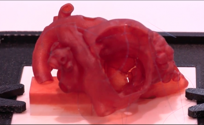
New 3-D Printing Innovations
About five years ago, 3-D printing began to make its appearance on the expo floor of RSNA. Today, there are numerous printer vendors exhibiting, several sessions on 3-D printing and 3-D printed anatomical models found in a large number of vendors’ booths promoting this technology. It has traditionally been a way to create models of complex or rare anatomy for use in clinical education. It has also seen a rapid uptake for planning and device sizing in complex surgical or interventional procedures. This is especially true in cardiology for structural heart procedures.
There were two new innovations in 3-D printing highlighted at RSNA this year. The first was the introduction of computer-aided design (CAD) software by 3-D printing vendor Materialise. An issue found with using 3-D printed anatomy is that it is static, such as the heart being frozen in one position of the cardiac cycle. So for planning purposes, printed models do not account for the dynamic nature of the anatomy expanding, contracting and twisting, which influence how an implanted device may behave inside a beating heart. The CAD software allows patient imaging datasets to be visualized from an engineering standpoint, where virtual devices can be implanted to show how they interact with the cardiac movement. This might help in designing better implantable devices or more accurate device sizing for transcatheter valves, septal occluders or left atrial appendage occluders.
Starting last fall, Materialise began its pre-market phase of development to validate its Mimics Innovation Suite CAD software for accurate 3-D modeling to help physicians plan complex transcatheter mitral valve replacement and repair (TMVR) procedures. One of its chief U.S. partners is Henry Ford Hospital in Detroit, where the software has been leveraged for better patient selection, procedural pre-planning and device sizing for off-label transcatheter mitral valve implantations. The hospital credits the software with improved outcomes. Henry Ford Hospital uses the software to create virtual 3-D anatomical models as the basis for assessing left ventricular outflow tract obstructions and other elements for planning complex mitral valve procedures.
Read the article “The Use of 3-D Printing in Cardiology,” which describes this software in more detail.
Watch the VIDEO “Use of 3-D Printing to Guide Structural Heart Interventions,” an interview with Dee Dee Wang, M.D., director of structural heart imaging at Henry Ford Hospital, and medical director, 3-D printing, Henry Ford Innovation Institute.
Another 3-D printing innovation unveiled at RSNA 2017 was the Biomimics product from Stratasys. It combines several existing 3-D printing materials with a new software that allows users to create custom, lifelike anatomy that mimics the physical traits of actual tissues. The company offers an on-demand printing service to create the models. These models can be used for education and to practice procedures. Stratasys demonstrated examples at RSNA, included suturing cardiac tissue and vessels, and implanting a spinal fixation bracket.
Interventional Imaging and Guidance Technologies
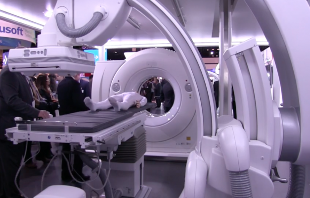
Siemens unveiled its new Nexaris therapy platform, which combines the vendor’s robotic angiography Artis pheno system with either an in-room CT system, or allows pairing with a Siemens MRI system.
The MRI configuration of the Nexaris system uses a removable angiography patient tabletop that detaches and rolls onto a special gurney. The patient can then be wheeled down to an MRI suite without the patient changing position. This allows for fused MRI-angio imaging. The two configurations are pending FDA clearance.
The CT version is a gantry on rails that can be moved up to the patient table, and the table slides into the gantry for CT imaging without moving the patient. The CT imaging can be used to create more detailed anatomical overlays on live angiography to aid procedural guidance, or to confirm a procedure is successful. Siemens said the system can save time and avoid the need for patient followup imaging.
Siemens said in an era of value-based reimbursements, hospitals need to confirm the treatment is successful before the patient gets off the table to avoid readmission or repeat procedures that they will not be reimbursed for.
GE Healthcare and cardiovascular imaging software provider Medis announced at the 2017 Transcatheter Cardiovascular Therapeutics (TCT) meeting a couple weeks prior to RSNA they are collaborating to add image-based fractional flow reserve (FFR) technology in GE’s angiography systems. The technology would eliminate the need for FFR pressure wires or the use of adenosine.
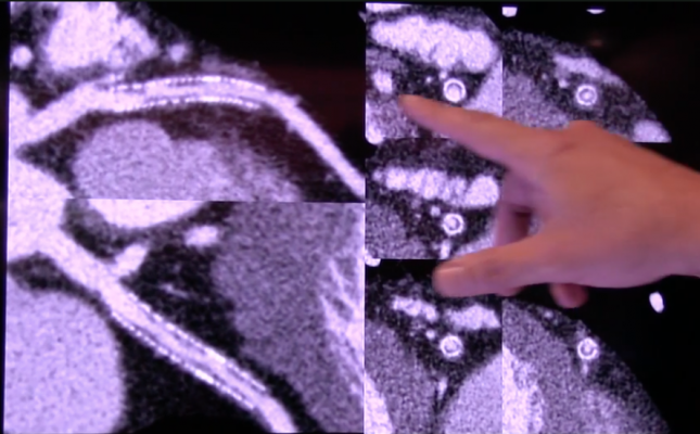
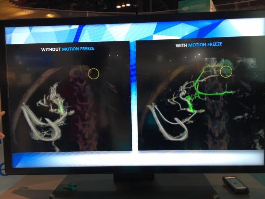
GE Healthcare also showed a cool new technology for its interventional guided therapy (IGS) angiography systems at the 2017 Radiological Society Of North America (RSNA) meeting. Rotational angiography 3-D CT-like imaging scans in the cath lab, are often suboptimal because of respiratory motion. A new Assist technology helps cancel out this motion to improve image quality. Critical areas of interest can sometimes be missed or obscured on images because of motion blur, but the new software helps preserved these areas by filtering out the motion.
Ultrasound Advances
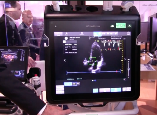
GE Healthcare rolled out its first implementation of artificial intelligence in its ultrasound systems, starting with its new point-of-care Venue system, designed for critical care, anesthesia and trauma. The user images the patient with a 3-D ultrasound probe and the system’s AI identifies the anatomy being imaged using anatomical landmarks. It then pulls out and displays the optimal views of that anatomy from the dataset. The idea is to save time and speed diagnosis by increasing the reproducibility of the standard views and measurements between various users, regardless of their level of training. GE said it plans to add this AI technology to all its ultrasound platforms in the coming years.
The GE AI technology is similar to Philips Healthcare’s Anatomical Intelligence used on its Epiq cardiac ultrasound platform. That technology uses 3-D dataset acquisitions of the heart to automatically identify all aspects of the cardiac anatomy and then display the ideal standard views. It then automatically performs quantification measurements. The Epiq platform does all this in seconds, greatly speeding exam times.
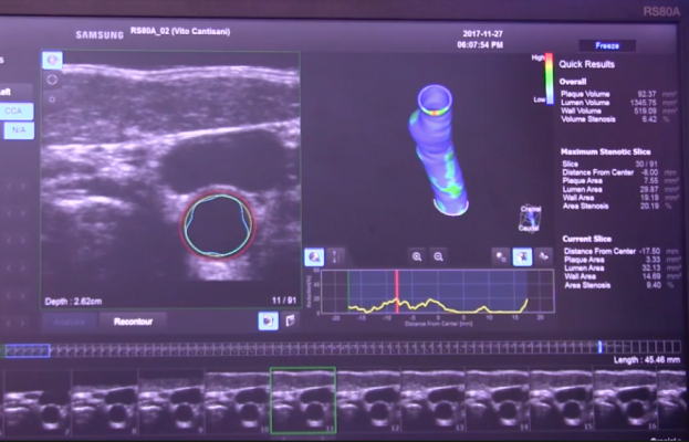
Samsung showed for the first time its RS80A ultrasound system’s new S3D Arterial Analysis package. It is used to evaluate the carotid artery for both overall cardiovascular risk and stroke risk. The technology goes way beyond the traditional carotid intima-media thickness (IMT) ultrasound risk assessments. The new Samsung software uses a 3-D ultrasound acquisition to reconstruct a 3-D image of the vessel showing the layers of the vessel wall and the plaque burden. It can be displayed like a CT dataset where the users can scroll through the slices of the vessel as a 3-D color-coded anatomical reconstruction, or in a virtual reconstruction of the vessel being split and unrolled as a flat plane. This last view is color coded as well to show areas of high and low plaque burden and may allow easier visualization of the plaque rather than viewing inside a cylinder-shaped vessel. The technology may offer a more detailed and accurate assessment of the carotids over traditional 2-D ultrasound views.
Read the realted article "Recent Advances in Echocardiography Technology"
MRI Advances
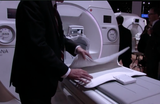
The most interesting magnetic resonance imaging advance at RSNA this year was GE Healthcare’s launch of its new AIR coil technology. It enables the creation of blanket-like MRI coils, rather than the hard plastic or semi-flexible coil sheets currently available. The AIR coils can be folded and bent to easily wrap around the patient’s body. The coils are extremely lightweight and use flexible electronics. The AIR technology also allows coils to overlap without cross-talk interference, unlike conventional coil technology.
GE, Philips and Siemens showed new software on their MRI systems than can reduce some exam times by a third or half of current time. This could help make cardiac MRI more attractive and economical than in the past.
One of the more practical new technologies we saw at RSNA 2017 was a smartphone app that allows simulation of scanning for patients with implanted medical devices to see if the implant will cause a patient safety issue. The user creates an avatar of their patient's sex, height, weight, etc., virtually implants the medical device they have and then runs a simulation of an MRI on the scanner they will be using to show where the magnetic fields are. The app turns the device red, green or yellow to show if it is in a dangerous field or pulse during the scan. It is very detailed and was fun to use. The app programmer also is an MRI tech, so they understood what is involved in scanning and safety issues related to implantable devices.
The invisible magnetic fields and RF energy pulses can have catastrophic effects on patients with medical metal implants. MRI expert Emanuel Kanal, M.D., University of Pittsburgh, developed the interactive app for iPhones to address the most common patient safety concerns involving implants. It includes a selection of various types of pacemakers, neurostimulators, stent grafts, spinal fixation devices and others. A report can be created with images to print or e-mail for the patient record. The app will be available from the Apple store starting in March 2018.
Watch "VIDEO: New App Improves MRI Safety For Implantable Devices"
Carotid Reporting Templates
Change Healthcare showed new vascular reporting templates for its cardiovascular information system, including one for stroke. Cardiac reporting systems started out a decade ago at first with basic coronary artery tree diagrams to illustrate stenting procedures or blockages on angiograms. Today vendors are expanding these diagrams, enabling them for more complex, interactive editing and expanding them to include all the major vasculature in the body.
Cardiology Related RSNA Study Presentations:
Male Triathletes May Be Putting Their Heart Health at Risk
CT Shows Enlarged Aortas in Former Pro Football Players
Fat Distribution in Women and Men Provides Clues to Heart Attack Risk
Other News and Video From RSNA 2017:
VIDEO: How Advanced Visualization and 3D Printing Can Improve Outcomes in Complex Cases
VIDEO: Key Imaging Technology Trends at RSNA 2017
VIDEO: Cybersecurity in the Medical Imaging Department
VIDEO: How Serious is MRI Gadolinium Retention in the Brain and Body?
VIDEO: Big Concerns Remain for MRI Gadolinium Contrast Safety at RSNA 2017
Radiology Has Failed to Properly Assess or Track MRI Gadolinium Contrast Safety
 May 12, 2020


