Nuclear imaging technology for both single photon emission computed tomography (SPECT) and positron emission tomography (PET) have made advancements in the past couple years. The main drivers for this have been a movement to digital imaging detectors to improve image quality and address radiation dose concerns, reimbursement and radiotracer supply issues. Other advancements have come in the areas of software to improve image reconstruction quality, offer better clinical qualification and analytics data.
“We have been practicing nuclear cardiology since the late 1970s, so people think of it as a stable and well-established modality, but if you look at the past five years, there has been a tremendous amount of advance in nuclear cardiological techniques,” said Prem Soman, M.D., Ph.D., director of nuclear cardiology at the Heart and Vascular Institute, University of Pittsburgh, and president of the American Society of Nuclear Cardiology (ASNC). “We have a whole new generation of SPECT cameras, we are expanding our imaging applications, we have made great strides in reducing our radiation dose, and PET is becoming more widely used, so I am very excited about the future of cardiac nuclear imaging.”
He explained one of the biggest advances in the last few years is the introduction of SPECT cameras using digital cadmium zinc telluride (CZT) detectors. These replace the old photomultiplier tubes (PMTs) that have been the industry standard for decades. CZT detectors are smaller to decrease the size of SPECT systems.
The biggest asset of CZT detectors is the improved image quality with direct digital transfer of photons into electrical signals to create images. The detectors are more sensitive that PMT-based cameras, so it enables exams with half the amount of radiotracers used in exams by conventional cameras. Alternatively, Soman said the usual dose can be used and the imaging time can be reduced by 50 percent, known as half-time imaging. The use of less radiotracer is also attractive from a supply standpoint, lowering operational costs and because there have been shortages of some SPECT radioisotopes over the past few years.
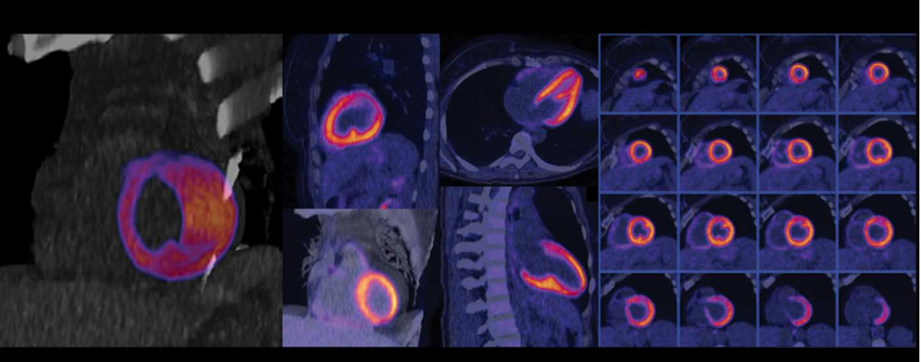
Soman also said hybrid systems that integrate a computed tomography (CT) scanner for PET-CT and SPECT-CT systems have become more popular in the market. These combined systems use CT for attenuation correct on the nuclear images, while adding CT anatomical image overlays improves the accuracy of diagnosis by visualizing the coronary anatomy to better pinpoint where blockages are causing perfusion defects.
Recent Nuclear Imaging System Introductions
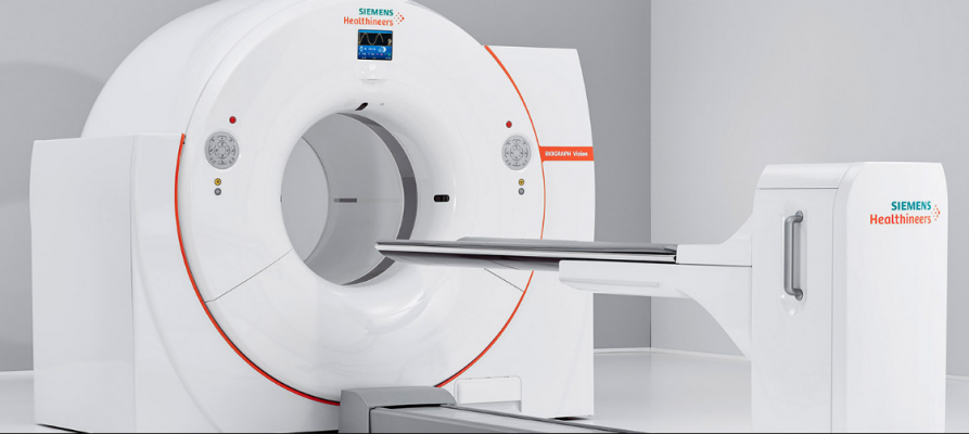
The most recent system to gain U.S. Food and Drug Administration (FDA) clearance was the Siemens Biograph Vision PET/CT in June, which features the company's new Optiso Ultra Dynamic Range (UDR) detector technology. It is based on silicon photomultipliers (SiPMs) rather than PMTs. The detector uses lutetium orthosilicate (LSO) crystal elements, which enable faster imaging and higher light output than commonly used bismuth germanate oxide (BGO) crystals. The LSO technology also offers higher image resolution, better image quality and enabling time-of-flight (TOF) acquisition. The size of LSO crystal elements, from 4 x 4 mm to 3.2 x 3.2 mm, results in higher spatial resolution and enables a smaller footprint for the system. With the smaller crystals and covering 100 percent of the area of the scintillator array with SiPMs, Siemens said the system can deliver the industry's fastest TOF, with a temporal resolution of 249 picoseconds. The Biograph Vision also has a large 78 cm bore to help reduce patient anxiety in tight spaces and fit larger bariatric patients.
The Siemens Biograph Horizon PET-CT system was cleared by the FDA in June 2017. It uses 4 mm LSO crystals that can visualize smaller details than previous-generation systems using the new detector design.
Siemens also debuted the Symbia Intevo Bold SPECT-CT system at the 2017 annual meeting of the Society of Nuclear Medicine and Molecular Imaging (SNMMI). The system addresses the growing trend of healthcare facilities using SPECT-CT in a dual-use setting, mirroring the rise of dual-use PET-CT. More facilities are also utilizing SPECT-CT systems as standalone or backup diagnostic CT systems. The system offers the SAFIRE (Sinogram Affirmed Iterative Reconstruction) algorithm to deliver excellent CT image quality while reducing patient radiation dose by as much as 60 percent. It also offers the iMAR (Iterative Metal Artifact Reduction) algorithm that reduces metal-related artifacts caused by metallic materials, such as orthopedic and dental implants.
In October 2016, GE Healthcare gained FDA clearance for its Discovery MI digital PET-CT system. It uses new detector technology that allows significantly better resolution. The LightBurst Digital Detector delivers up to two times improvement in volumetric resolution. GE also said it has the highest National Electrical Manufacturers Association (NEMA) sensitivity of any time-of-flight PET system. These expanded capabilities enable up to a 50 percent reduction in tracer usage without impacting image quality.
“One of the most striking features of Discovery MI is the improved sensitivity,” said Ronny Ralf Buechel, M.D., a cardiologist and nuclear medicine physician from Zurich. “We didn’t know how low we could go with the injected activity and radiation dose, so we began to cautiously lower the dose. Now, we are around 50 percent of the injected dose for PET scans compared to the prior system — around 1 mSv for a complete rest/stress study, and what remains important for cardiac imagers is the reduced activity of ammonia without any deterioration in image quality.”
The CT portion of the Discovery MI delivers 100 percent better spatial resolution than previous GE systems with no increase in image noise. This is partly enabled by the systems ASiR-V iterative reconstruction and Smart Metal Artifact Reduction (MAR) software.
GE’s Discovery NM/CT 670 CZT SPECT-CT system, introduced in 2016, debuted cadmium zinc telluride (CZT) digital detector technology, which greatly improves image quality over the old industry standard PMTs. CZT technology allows for direct conversion of photons into a digital signal, which makes the image transfer more efficient. This allows for half-time imaging or the ability to significantly reduce radiotracer dose.
Research collaborations with Rambam Health Center, Barnes-Jewish Hospital, St. Jean Clinic and Hospices Civils de Lyon have demonstrated the Discovery NM/CT 670 CZT technology helped lead to a 75 percent dose reduction, or time reduction in exams.
GE Healthcare recently introduced the Discovery 670 DR, a digital-ready conventional SPECT/CT system requiring a simple two-day in-field upgrade to CZT technology. The system’s modular design offers easy upgradability to digital. Both Discovery NM/CT 670 CZT and Discovery 670 DR are now powered by GE’s latest 16-slice CT system, offering additional improvements in CT image quality with an overlap reconstruction which enables 32 slices per rotation.
At the 2017 Society of Nuclear Medicine and Molecular Imaging (SNMMI) meeting, GE Healthcare also introduced the Discovery MI DR PET-CT system. It uses PMT technology, but is built with a modular design to allow customers to cost-effectively upgrade later to digital detectors.
Philips Healthcare's Vereos Digital PET/CT is a fully digital system featuring digital photon counting (DPC) technology. It offers improved detectability and characterization of small lesions in oncology. The company said the system provides uncompromised detectability and quantification at half the PET dose. Vereos also provides lesion detectability in one-tenth of the time.
The Philips Ingenuity TF PET/CT uses the vendor’s Astonish TOF technology for enhanced image contrast, resolution and quality. The company said it improves contrast by up to 30 percent compared to non-TOF systems. It also improves signal-to-noise ratio, resulting in exceptional image quality, higher speed and enhanced accuracy. The CT system uses iPatient, iDose⁴ and metal artifact reduction for implants (O-MAR) software to improve the CT image quality and lower the X-ray radiation exposure.
The Canon Medical Systems Celesteion PureVision Edition PET/CT system delivers the largest PET/CT bores and field of view to help enhance patient comfort, the company said. The system has a 70 cm PET and CT field of view (FOV), compared to the smaller 50 cm FOV on some systems. The CT system offers a 90 cm wide bore. The PET system has an 88 cm bore. The PET system also provides 394 ps TOF, a 19.6 cm axial FOV and uses 4 x 4 mm detector crystals.
Information Technology Now Plays a Key Role in Nuclear Imaging
Nuclear medicine technology has significantly evolved in recent years, but in more recent times this has branched out to not only include scanner advancements, but also advanced informatics, quantification software and analytics. As value-based care changes the way healthcare organizations approach care delivery, there is a greater need for easy-to-use, fast and precise imaging. With increased access to better data, clinicians are looking for ways to make that data actionable.This includes customizing patient treatments and better management of imaging departments.
Buyers for these newer systems should be looking at the software to help enhance image quality, aid or automate quantification and measurements, help monitor and reduce tracer usage or dose, and offer analytics on procedures, operator statistics and information on scanner performance. This level of analytical information can help better manage the department, help identify workflow bottlenecks and performance issues, and help lower dose.
For example, the Symbia Intevo Bold SPECT-CT system uses the xSPECT Quant quantification technology. This enables automated, accurate and reproducible quantification of not only technetium-99m, but also iodine-123, lutetium-177 and indium-111. This capability extends the use of advanced SPECT quantification from general nuclear medicine and bone studies to indications including neurological disorders, neuroendocrine tumors and prostate cancer.
Related Nuclear Cardiology Content:
New ASNC SPECT Imaging Guideline Addresses Advances in Myocardial Perfusion Imaging
Dose-Lowering Practices for Nuclear Cardiology
The Past, Present and Future of PET/MRI Scanners
Recent Advances in Cardiac Nuclear Imaging Technology
VIDEO: PET vs. SPECT in Nuclear Cardiology and Recent Advances in Technology — An interview with Prem Soman, M.D.
VIDEO: Trends in Nuclear Cardiology Imaging — A discussion with David Wolinsky, M.D., director of nuclear cardiology at Cleveland Clinic Florida and past-president of the ASNC

 January 05, 2023


 imaging program. She spoke on this topic at the 2019 meeting of the American Society Nuclear Cardiology (ASNC) and led a tour with attendees of the PET-CT system at Rush, which was located close to the conference. #ASNC")