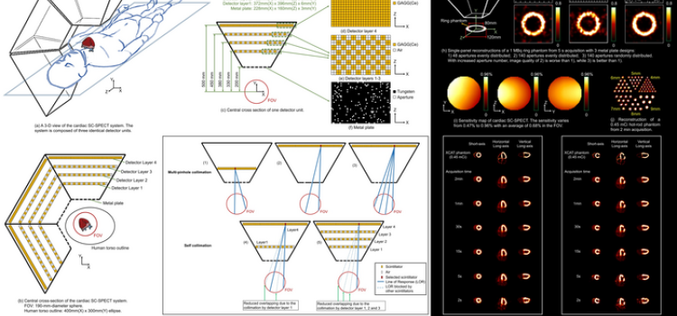
June 15, 2022 — A cardiac SPECT imaging system performs scans 10 to 100 times faster than current SPECT systems, according to new research presented at the Society of Nuclear Medicine and Molecular ...
SPECT Imaging
Single-photon emission computerized tomography (SPECT) imaging is a nuclear imaging technology (also referred to as molecular imaging) that produces images showing how organs work. The is eanbled by the use of radiotracers, usually attached to sugars. The cells in the body metabolize the sugar and the nuclear images show areas of high and low sugar uptake. This allows imaging of ischemia or infarct in the heart or other organs, or areas of high sugar uptake caused by cancers, which usually have a much higher metabolism than health cells.
-
-
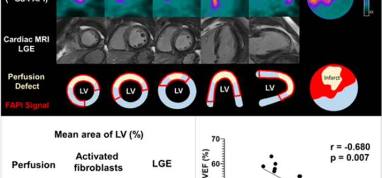
June 15, 2022 — Poor functional outcomes after a heart attack can be predicted with a new PET imaging agent, 68Ga-FAPI-46. According to research presented at the Society of Nuclear Medicine and ...
-

American Society of Nuclear Cardiology (ASNC) President Dennis Calnon, M.D., MASNC, FASE, FSCCT, director of cardiac imaging for McConnell Heart Hospital at Riverside Methodist Hospital, and director ...
-

American Society of Nuclear Cardiology (ASNC) President Dennis Calnon, M.D., MASNC, FASE, FSCCT, director of cardiac imaging for McConnell Heart Hospital at Riverside Methodist Hospital, and director ...

Sponsored Content — According to the American Heart Association, cardiovascular disease is the leading cause of death in ...
 March 13, 2026
March 28, 2024 — Cleerly, the company on a mission to create a new standard of care to aid in the diagnosis of heart ...
March 28, 2024
January 3, 2024 — HeartFlow, Inc., a leader in non-invasive artificial intelligence (AI) precision coronary care ...
January 03, 2024Sponsored Content

Feature | Cardiac Imaging
Sponsored Content — According to the American Heart Association, cardiovascular disease is the leading cause of death in ...
March 13, 2026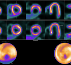
June 28, 2023 — A novel PET perfusion radiotracer, 18F-flurpiridaz, can diagnose coronary artery disease (CAD) in obese ...
June 28, 2023 June 05, 2023
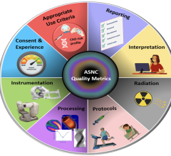
January 13, 2023 — Nuclear stress testing performed with single-photon emission computed tomography (SPECT) is the most ...
January 13, 2023
January 5, 2023 — StreamlineMD has provided updates for 2023 Radiology and IR CPT coding, which may impact your practice ...
January 05, 2023
October 31, 2022 — Several recent discoveries show that the accuracy of diagnosing coronary artery disease and ...
October 31, 2022
September 12, 2022 — Royal Philips, a global leader in health technology, announced new milestones in the development of ...
September 12, 2022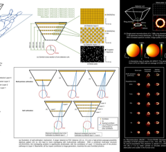
June 15, 2022 — A cardiac SPECT imaging system performs scans 10 to 100 times faster than current SPECT systems ...
June 15, 2022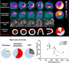
June 15, 2022 — Poor functional outcomes after a heart attack can be predicted with a new PET imaging agent, 68Ga-FAPI ...
June 15, 2022
February 18, 2022 — GE Healthcare has announced that it has received approval from the European Medicines Agency (EMA) ...
February 18, 2022
American Society of Nuclear Cardiology (ASNC) President Dennis Calnon, M.D., MASNC, FASE, FSCCT, director of cardiac ...
February 01, 2022
American Society of Nuclear Cardiology (ASNC) President Dennis Calnon, M.D., MASNC, FASE, FSCCT, director of cardiac ...
February 01, 2022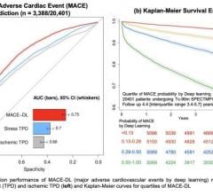
June 14, 2021 — An advanced artificial intelligence technique known as deep learning can predict major adverse cardiac ...
June 14, 2021