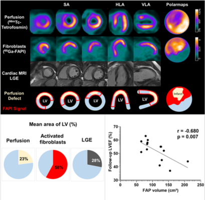
Representative case with acute anterior wall myocardial infarction: Myocardial perfusion images using 99mTc-tetrofosmin at rest (first row), 68Ga-FAPI-46 PET (second row), late gadolinium enhancement (LGE) from cardiac magnetic resonance (third row) and schematic drawings of the left ventricle (fourth row). Area of fibroblast activation exceeds infarct area and LGE signal, most common type of myocardial FAP-distribution. Bottom left: Mean areas of perfusion defect, FAPI-signal and LGE of 35 studied patients. Bottom right: Global myocardial FAP volume early after acute myocardial infarction inversely correlates with left ventricular ejection fraction (LV-EF) at follow-up in the chronic stage (n=14 patients). Image courtesy of Johanna Diekmann, James Thackeray, Thorsten Derlin, Christoph Czerner, Tobias Ross, and Frank Bengel, Department of Nuclear Medicine, Hannover Medical School, Hannover, Niedersachsen, Germany; and Tobias Koenig, Jonas Neuser, Andreas Schaefer, Jochen Tillmans, and Johann Bauersachs, Department of Cardiology and Angiology, Hannover School of Medicine, Hannover, Niedersachsen, Germany.
June 15, 2022 — Poor functional outcomes after a heart attack can be predicted with a new PET imaging agent, 68Ga-FAPI-46. According to research presented at the Society of Nuclear Medicine and Molecular Imaging 2022 Annual Meeting, when correlated with cardiac MRI, 68Ga-FAPI-46 PET is new specific indicator of active fibrosis and can identify a patient’s chances of recovery. The detailed depiction of 68Ga-FAPI-46 PET signal after acute anterior myocardial infarction has been selected as SNMMI’s 2022 Image of the Year.
Each year, SNMMI chooses an image that best exemplifies the most promising advances in the field of nuclear medicine and molecular imaging. The state-of-the-art technologies captured in these images demonstrate the capacity to improve patient care by detecting disease, aiding diagnosis, improving clinical confidence, and providing a means of selecting appropriate treatments. This year, the SNMMI Henry N. Wagner, Jr., Image of the Year was chosen from more than 1,000 abstracts submitted to the meeting and voted on by reviewers and the society leadership.
Acute myocardial infarction, commonly known as heart attack, is one of the leading causes of death worldwide. After a heart attack, the heart goes through a series of changes at the molecular and cellular level. The fibroblast activation protein (FAP) is overexpressed as these changes occur and can provide useful information about a patient’s prognosis.
“Molecular PET imaging of the fibroblast activation protein has recently been evaluated in patients after acute myocardial infarction,” stated Johanna Diekmann, MD, clinician scientist in the Department of Nuclear Medicine at the Hannover School of Medicine in Hannover, Germany. “In our study, we sought to obtain further insights by correlating FAP-targeted PET imaging with tissue characteristics from cardiac MRI, as well as functional outcome.”
Thirty-five patients underwent 68Ga-FAPI-46 PET/CT, perfusion SPECT, and cardiac MRI within 11 days after a heart attack. Cardiac FAP-volume was determined by PET imaging and infarct size defined by SPECT imaging. Cardiac MRI showed functional parameters, area of injury and tissue mapping. These datapoints were then compiled to examine potential correlations.
In all patients the FAP-upregulation was significantly larger than both the infarct size and the area of injury as defined by SPECT and cardiac MRI, respectively. A higher extent of myocardial FAP upregulation was predictive of subsequent left ventricular dysfunction. As such, the authors concluded, fibroblast activation in non-infarcted areas of the heart may contribute to adverse outcomes.
“Myocardial infarction is an important contributor to the development of heart failure, but the early molecular processes involved in the transition from initial injury to heart failure are under-treated,” said Diekmann. “New antifibrotic therapies (such as CAR-T cell therapies) could significantly change future therapy of heart failure. Using FAPI-PET to select patients suitable for therapy would open a new major application for PET in fibrosis and cardiac diseases.”
“FAPI is a very exciting radiotracer that holds great potential for the future of nuclear medicine and molecular imaging,” noted SNMMI Scientific Program chair Heather Jacene, MD. “As shown in the Image of the Year, 68Ga-FAPI-46 PET/CT clearly indicates pro-fibrotic activity after acute myocardial infarction. The predictive value of FAPI-PET for the magnitude of subsequent decline in global cardiac function, if further validated, may be used in the future to help select the most suitable patients for anti-fibrotic therapies which are currently under development. This could ultimately have a powerful impact on cardiovascular medicine.”
Abstract 413. “Predicting Remodeling and Outcome from Molecular Imaging of Fibroblast Activation in Patients after Acute Myocardial Infarction,” Johanna Diekmann, James Thackeray, Thorsten Derlin, Christoph Czerner, Tobias Ross, and Frank Bengel, Department of Nuclear Medicine, Hannover Medical School, Hannover, Niedersachsen, Germany; and Tobias Koenig, Jonas Neuser, Andreas Schaefer, Jochen Tillmans, and Johann Bauersachs, Department of Cardiology and Angiology, Hannover School of Medicine, Hannover, Niedersachsen, Germany.
For more information: www.snmmi.org
 June 28, 2023