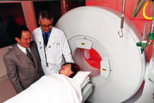
A team led by UTHealth Houston's K. Lance Gould, MD, (center) used PET imaging technology (shown above) to map coronary blood flow and its outcomes among heart disease patients. (Photo by UTHealth Houston)
December 1, 2022 — A new method for determining whether patients with heart disease need coronary stents or bypass surgery is more effective than the angiogram, which is currently used, according to research from UTHealth Houston Heart & Vascular.
A team led by K. Lance Gould, MD, professor and the Martin Bucksbaum Distinguished University Chair in Heart Disease with McGovern Medical School at UTHealth Houston, used positron emission tomography (PET) imaging technology to map coronary blood flow and its outcomes – namely, subendocardial ischemia – among patients with heart disease. The study was published in JACC Journals.
Most forms of heart disease cause myocardial damage. Myocardial ischemia occurs when blood flow to the heart is reduced, preventing the heart muscle from receiving enough oxygen. When myocardial ischemia affects the deep, or subendocardial, layer of the left ventricular muscle, it is known as subendocardial ischemia.
Subendocardial ischemia is commonly diagnosed in patients with cardiovascular disease, but it is not quantified by current imaging tools. Gould’s team developed the PET technology, software, and clinical validation for defining the size and severity of this early stage of coronary artery disease.
“The cumulative data reveals that all randomized trials of coronary stents and bypass surgery have failed to improve survival after revascularization due to profoundly flawed patient selection based on the angiogram,” said Gould, who was first author on the study. “Thus, the coronary angiogram is not the gold standard for determining stents or bypass surgery, but rather, quantitative myocardial perfusion by PET is the gold standard.”
Significantly, the paper confirmed the team’s previous research by proving the PET threshold of severity at which stents and bypass surgery improve survival compared to medical treatment alone. The angiogram – an X-ray test that helps doctors evaluate blockages in the arterial system – shows how to do stents or bypass surgery, Gould said, but not whether those procedures should be done at all.
Gould, who began working at UTHealth Houston in 1979 as a professor and director of the division of cardiology, stepped aside from administrative duties in 1987 to focus clinically and scientifically on PET imaging and quantitative coronary arteriography for identifying segmental and diffuse coronary artery disease, measuring its severity and reversing it by vigorous risk factor modification.
“Several equally paradigm-changing papers are underway for the coming year,” Gould said. “For example, our preliminary data show that virtual revascularization on cardiac PET images predicts survival outcomes before actually doing stents or bypass surgery, as a guide to making decisions for or against those procedures.”
Nils P. Johnson, MD, MS, professor and Weatherhead Distinguished Chair of Heart Disease with McGovern Medical School, was the study’s senior author. Other co-authors from the Division of Cardiovascular Medicine at McGovern Medical School included Tung Nguyen, BS; Richard Kirkeeide, PhD; Amanda E. Roby, CNMT, RT(N); Linh Bui, MD; Danai Kitkungvan, MD; Monica B. Patel, MD; Mohammad Madjid, MD; and Mary Haynie, RN, MBA. Co-authors with UTHealth Houston School of Public Health included Dejian Lai, PhD, and Ruosha Li, PhD. Jagat Narula, MD, PhD, with Mount Sinai Heart at Mount Sinai Morningside and Icahn School of Medicine at Mount Sinai in New York, also contributed to the study.
For more information: https://www.uth.edu/
 November 24, 2025