September 7, 2022 — The American Society of Nuclear Cardiology (ASNC) and three partner societies have come together to answer cardiac imagers’ questions about SPECT/CT and PET/CT imaging with ...
ASNC
This channel contains news about the American Society of Nuclear Cardiology (ASNC), including coverage of its annual meeting and links to recently released practice guidelines. ASNC is a leading resource for the subspecialty of cardiac nuclear imaging (also called molecular imaging). Cardiac imaging with either PET or SPECT is primarily used for myocardial perfusion imaging (MPI), which shows area of areas of reduced blood flow due to ischemia or infarct.
-
-
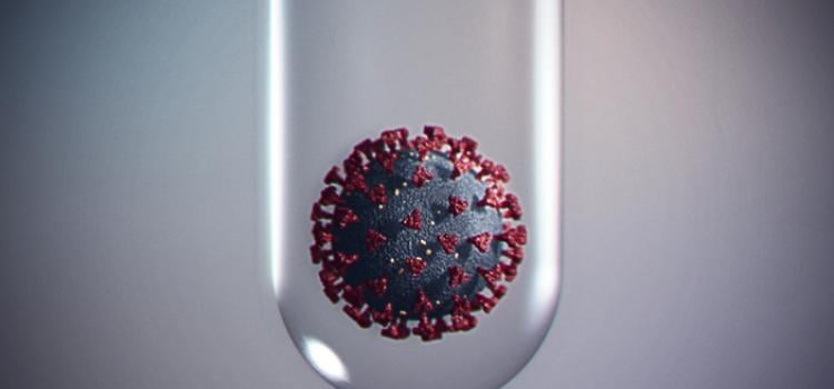
May 4, 2022 — While the COVID-19 pandemic transitions through various phases, nuclear cardiology teams should maintain a flexible approach about which testing protocols to follow in different clinical ...
-
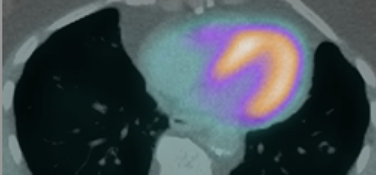
 April 21, 2022 – Coronary artery disease (CAD) is one of the major causes of mortality and morbidity worldwide. Noninvasive imaging modalities play a fundamental role in the evaluation and management ...
 -
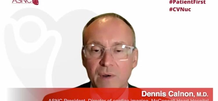
American Society of Nuclear Cardiology (ASNC) President Dennis Calnon, M.D., MASNC, FASE, FSCCT, director of cardiac imaging for McConnell Heart Hospital at Riverside Methodist Hospital, and director ...

Sept. 18, 2025 — The Society of Nuclear Medicine and Molecular Imaging (SNMMI), American Society of Nuclear Cardiology ...
 September 22, 2025
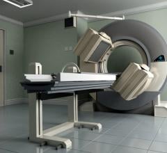 June 05, 2023
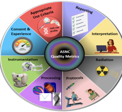
January 13, 2023 — Nuclear stress testing performed with single-photon emission computed tomography (SPECT) is the most ...
January 13, 2023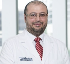
January 5, 2023 — Mouaz H. Al-Mallah, MD, MSc, FASNC, director of the cardiovascular PET program at the Houston ...
January 05, 2023
September 7, 2022 — The American Society of Nuclear Cardiology (ASNC) and three partner societies have come together to ...
September 07, 2022
August 25, 2022 — The results of “A Phase 3, Open-label, Multicenter Study of Flurpiridaz (F18) Injection for Positron ...
August 25, 2022
August 12, 2022 — The results of “A Phase 3, Open-label, Multicenter Study of Flurpiridaz (F18) Injection for Positron ...
August 12, 2022
August 10, 2022 — On July 27, ASNC joined a coalition of more than 100 medical societies urging Congress to pass ...
August 10, 2022
July 26, 2022 — ASNC Past President Sharmila Dorbala, MD, MPH, MASNC, will present the Mario Verani Memorial Lecture ...
July 26, 2022
July 19, 2022 — On July 7, the Centers for Medicare & Medicaid Services (CMS) released the 2023 Medicare Physician Fee ...
July 19, 2022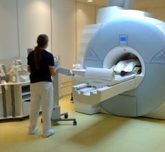
July 11, 2022 – The American Society of Nuclear Cardiology (ASNC) and the Society of Nuclear Medicine and Molecular ...
July 11, 2022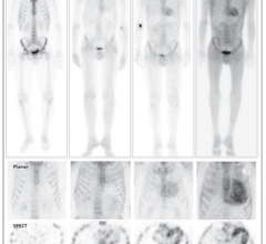
June 24, 2022 — Shortages of pyrophosphate (PYP), the radiopharmaceutical most commonly used in the U.S. for noninvasive ...
June 24, 2022
May 4, 2022 — While the COVID-19 pandemic transitions through various phases, nuclear cardiology teams should maintain a ...
May 04, 2022
April 21, 2022 – Coronary artery disease (CAD) is one of the major causes of mortality and morbidity worldwide ...
April 21, 2022April 13, 2022 — The Society of Nuclear Medicine and Molecular Imaging (SNMMI) and the American Society of Nuclear ...
April 13, 2022