Nov. 17, 2025 — GE HealthCare has announced that Cardiovascular Associates of America (CVAUSA) plans to broaden its adoption of GE HealthCare’s FDA-approved cardiac positron emission tomography (PET) ...
PET Imaging
Positron emission tomography (PET) is a nuclear imaging technology (also referred to as molecular imaging) that enables visualization of metabolic processes in the body. The basics of PET imaging is that the technique detects pairs of gamma rays emitted indirectly by a positron-emitting radionuclide (also called radiopharmaceuticals, radionuclides or radiotracer). The tracer is injected into a vein on a biologically active molecule, usually a sugar that is used for cellular energy. PET systems have sensitive detector panels to capture gamma ray emissions from inside the body and use software to plot to triangulate the source of the emissions, creating 3-D computed tomography images of the tracer concentrations within the body.
-

-

July 21, 2025 — Long COVID patients with abnormal cardiopulmonary PET/MR findings may be more likely to develop heart and lung diseases, according to new research published in the July issue of The ...
-
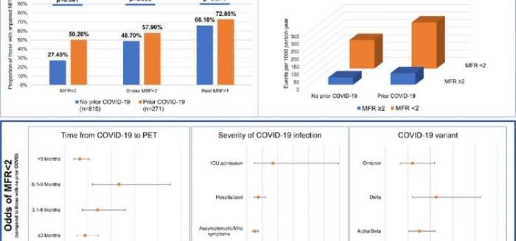
June 9, 2023 — Patients infected with beta and delta COVID-19 variants, and those who required hospital stays for COVID-19 infection, were more likely to experience heart issues associated with long ...
-
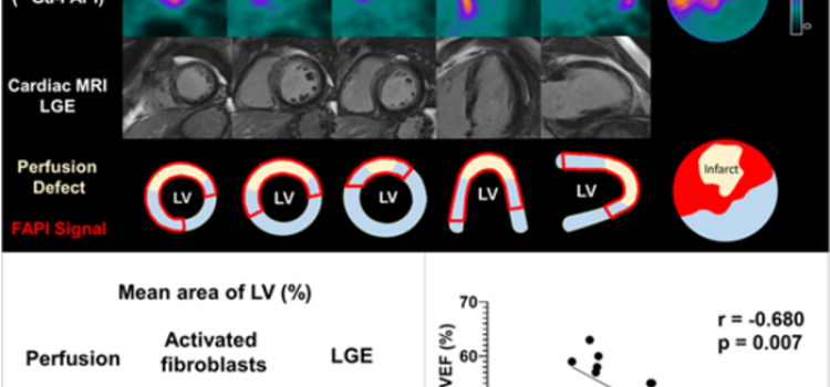
June 15, 2022 — Poor functional outcomes after a heart attack can be predicted with a new PET imaging agent, 68Ga-FAPI-46. According to research presented at the Society of Nuclear Medicine and ...

May 7, 2026 — Bayer has announced positive topline results from the Phase III REVEAL study, an investigator-initiated ...
 May 07, 2026
Nov. 17, 2025 — GE HealthCare has announced that Cardiovascular Associates of America (CVAUSA) plans to broaden its ...
November 17, 2025
Sept. 18, 2025 — The Society of Nuclear Medicine and Molecular Imaging (SNMMI), American Society of Nuclear Cardiology ...
September 22, 2025Sponsored Content

Feature | Cardiac Imaging
Cardiac positron emission tomography (PET) is growing in popularity among cardiologists because it provides the ability ...
March 05, 2024
Sept. 8, 2025 — GE HealthCare recently announced a Distribution and Services Agreement (DSA) with CardioNavix, a part of ...
September 08, 2025
July 21, 2025 — Long COVID patients with abnormal cardiopulmonary PET/MR findings may be more likely to develop heart ...
July 22, 2025
Feb. 25, 2025— GE HealthCare has delivered the first patient doses of Flyrcado (flurpiridaz F 18) injection, a unit dose ...
March 03, 2025
July 25, 2024 — Positron Corporation, a leading molecular imaging medical device company offering PET & PET-CT imaging ...
July 25, 2024
June 18, 2024 — Positron Corporation, a leading molecular imaging medical device company offering PET and PET-CT ...
June 18, 2024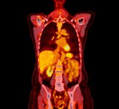
June 14, 2024 — Positron Corporation, a leading molecular imaging medical device company offering PET and PET-CT(Positro ...
June 14, 2024
Cardiac positron emission tomography (PET) is growing in popularity among cardiologists because it provides the ability ...
March 05, 2024
January 3, 2024 — HeartFlow, Inc., a leader in non-invasive artificial intelligence (AI) precision coronary care ...
January 03, 2024
November 21, 2023 — Heuron, a medical AI (artificial intelligence) imaging software solution company, under the ...
November 21, 2023
October 5, 2023 — Jubilant DraxImage Inc., a wholly-owned subsidiary of Jubilant Pharma Limited, has entered into an ...
October 05, 2023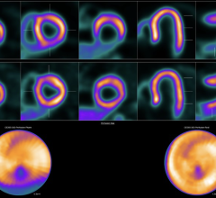
June 28, 2023 — A novel PET perfusion radiotracer, 18F-flurpiridaz, can diagnose coronary artery disease (CAD) in obese ...
June 28, 2023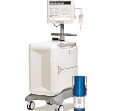
June 26, 2023 — Jubilant DraxImage Inc., dba Jubilant Radiopharma, a wholly-owned subsidiary of Jubilant Pharma Limited ...
June 26, 2023