September 12, 2012 – Positron Corp. announced that Jubilant DraxImage Inc., a Jubilant Life Sciences company, and ...
PET Imaging
Positron emission tomography (PET) is a nuclear imaging technology (also referred to as molecular imaging) that enables visualization of metabolic processes in the body. The basics of PET imaging is that the technique detects pairs of gamma rays emitted indirectly by a positron-emitting radionuclide (also called radiopharmaceuticals, radionuclides or radiotracer). The tracer is injected into a vein on a biologically active molecule, usually a sugar that is used for cellular energy. PET systems have sensitive detector panels to capture gamma ray emissions from inside the body and use software to plot to triangulate the source of the emissions, creating 3-D computed tomography images of the tracer concentrations within the body.
 September 12, 2012
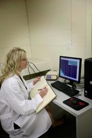
August 27, 2012 — Positron Corp., a molecular imaging healthcare company, announced the submission of a drug master file ...
August 27, 2012August 21, 2012 — The U.S. Food and Drug Administration (FDA) granted marketing approval for cfrQuant, coronary flow ...
August 21, 2012Sponsored Content

Feature | Cardiac Imaging
Cardiac positron emission tomography (PET) is growing in popularity among cardiologists because it provides the ability ...
March 05, 2024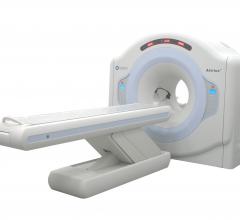
August 20, 2012 — Positron Corp. announced the use of its Attrius system at Caring Heart and Brain Imaging Inc. of ...
August 20, 2012August 14, 2012 — Positron Corp. announced the transfer, consolidation and integration of its radiopharmaceutical operat ...
August 14, 2012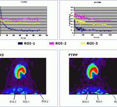
August 3, 2012 — FluoroPharma Medical Inc., a company specializing in the development of novel diagnostic imaging ...
August 03, 2012July 25, 2012 — Positron Corp. announced approval of a pledge for $15 million in tax increment financing (TIF) bonds fro ...
July 25, 2012July 13, 2012 — Royal Philips Electronics announced that it has embarked on a five-year joint research and development ...
July 13, 2012
June 8, 2012 — At this year’s SNM Annual Meeting, June 9-13, Philips Healthcare is highlighting its portfolio of nuclear ...
June 08, 2012May 18, 2012 - New research from the University at Buffalo suggests that cardiologists may have a new way to identify ...
May 18, 2012April 27, 2012 — More than 150 molecular imaging professionals gathered last week at the Society of Nuclear Medicine’s ...
April 27, 2012April 9, 2012 — After witnessing a downtrend for several years, revenues in the U.S. nuclear medicine and positron ...
April 09, 2012April 6, 2012 — Positron Corp. announced the approval of its Nuclear Regulatory Commission (NRC) manufacturing and ...
April 06, 2012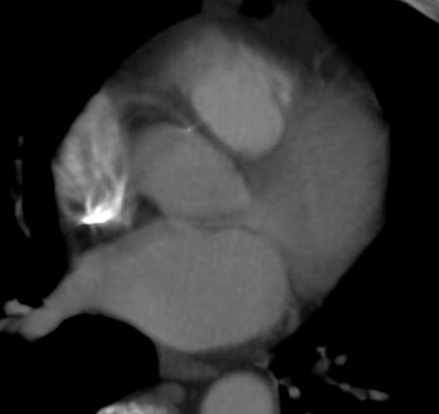
March 22, 2012 – Addressing concerns over radiation from cardiac imaging and procedures, the American College of ...
March 22, 2012
January 30, 2012 — Positron Corp., a molecular imaging healthcare company specializing in the field of nuclear ...
January 30, 2012