The fingerprints of value-added medicine were all over products and works-in-progress on the exhibit floor of the annual ...
PET Imaging
Positron emission tomography (PET) is a nuclear imaging technology (also referred to as molecular imaging) that enables visualization of metabolic processes in the body. The basics of PET imaging is that the technique detects pairs of gamma rays emitted indirectly by a positron-emitting radionuclide (also called radiopharmaceuticals, radionuclides or radiotracer). The tracer is injected into a vein on a biologically active molecule, usually a sugar that is used for cellular energy. PET systems have sensitive detector panels to capture gamma ray emissions from inside the body and use software to plot to triangulate the source of the emissions, creating 3-D computed tomography images of the tracer concentrations within the body.
 March 27, 2019
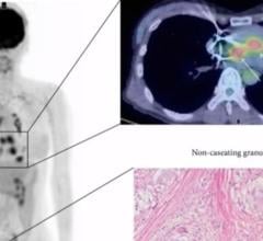
Raza Alvi, M.D., a research fellow in radiology at Massachusetts General Hospital, has been involved in a study of a ...
March 22, 2019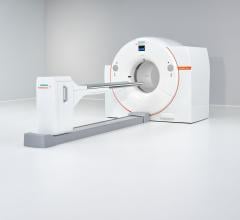
March 5, 2019 — Siemens Healthineers’ new Biograph Vision positron emission tomography/computed tomography (PET/CT) syst ...
March 06, 2019Sponsored Content

Feature | Cardiac Imaging
Cardiac positron emission tomography (PET) is growing in popularity among cardiologists because it provides the ability ...
March 05, 2024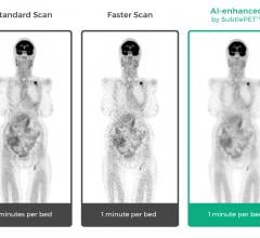
December 5, 2018 — Subtle Medical announced 510(k) clearance from the U.S. Food and Drug Administration (FDA) to market ...
December 05, 2018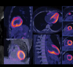
September 27, 2018 — Advancements in healthcare technology, particularly in the surgery category, have led to an ...
September 27, 2018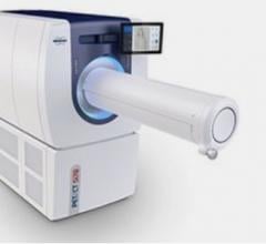
September 26, 2018 — Bruker recently announced the introduction of the new preclinical PET/CT Si78 scanner for whole ...
September 26, 2018
Robert C. Hendel, M.D., FACC, FAHA, MASNC, director, Tulane Heart and Vascular Institute, explains the impact the ASNC h ...
July 25, 2018
July 24, 2018 — In celebration of its 25th anniversary, the Journal of Nuclear Cardiology (JNC) is highlighting the top ...
July 24, 2018
July 17, 2018 — A new way to examine stress and inflammation in the heart will help Parkinson’s researchers test new ...
July 17, 2018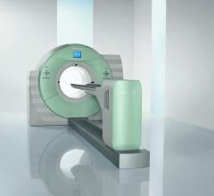
June 21, 2018 — Siemens Healthineers will announce U.S. Food and Drug Administration (FDA) clearance of four new system ...
June 21, 2018
Feature | Nuclear Imaging | Dave Fornell, Editor
Nuclear imaging technology for both single photon emission computed tomography (SPECT) and positron emission tomography ...
June 15, 2018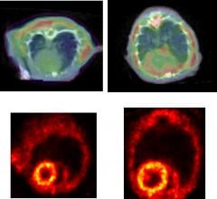
May 11, 2018 — A team led by Johns Hopkins University Researchers has discovered that protein clumps appear to ...
May 11, 2018
Feature | Nuclear Imaging | Dave Fornell
Cardiac nuclear myocardial perfusion imaging (MPI) has been a mature area of imaging for years, but has recently started ...
September 19, 2017
Prem Soman, M.D., director of nuclear cardiology at the Heart and Vascular Institute, University of Pittsburgh, and ...
August 24, 2017
Randall Thompson, M.D., outlines three new CPT codes for FFR-CT, a smart phone-based single-lead ECG system and PET ...
July 13, 2017