Nov. 17, 2025 — GE HealthCare has announced that Cardiovascular Associates of America (CVAUSA) plans to broaden its adoption of GE HealthCare’s FDA-approved cardiac positron emission tomography (PET) ...
PET-CT
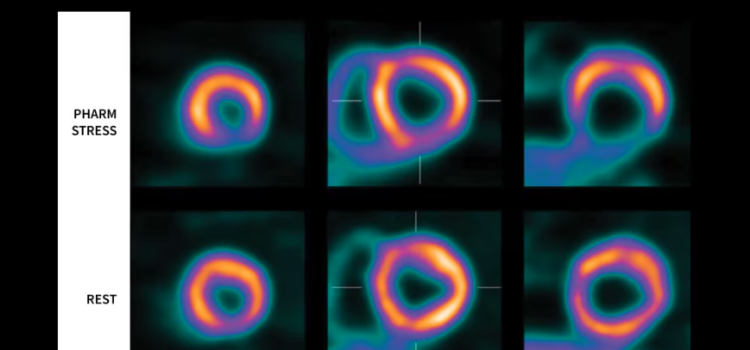
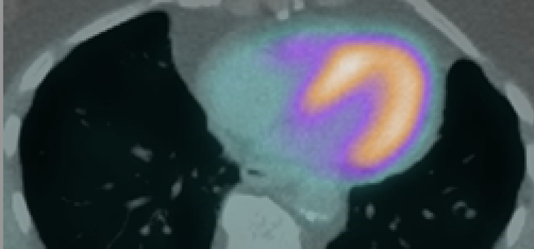
PET-CT combines positron emission tomography (PET) detectors and computed tomography (CT) into one imaging system. PET requires the use of CT for image attenuation correction. The CT scanners that can be installed on these systems include 4-slice, up to workhorse 64-slice or higher systems. High slice CT is usually an option picked by hospitals that plan to use the PET-CT scanner for standard CT only imaging exams as well. The CT scan anatomical imaging can be fused with the nuclear PET imaging to show anatomical landmarks. The CT component can also be used in cardiac PET to perform a coronary calcium scoring exam to offer a risk assessment for future heart attack risk.
-

-
June 23, 2025 — GE HealthCare’s commitment to advancing precision care in cardiology through its molecular imaging solutions will be on display at the Society of Nuclear Medicine and Molecular Imaging ...
-
September 7, 2022 — The American Society of Nuclear Cardiology (ASNC) and three partner societies have come together to answer cardiac imagers’ questions about SPECT/CT and PET/CT imaging with ...
-
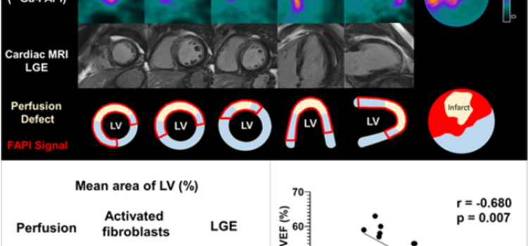
June 15, 2022 — Poor functional outcomes after a heart attack can be predicted with a new PET imaging agent, 68Ga-FAPI-46. According to research presented at the Society of Nuclear Medicine and ...

March 17, 2026 — Stern Cardiovascular and Atria Health have announced a strategic partnership that will accelerate ...
 March 17, 2026
Nov. 17, 2025 — GE HealthCare has announced that Cardiovascular Associates of America (CVAUSA) plans to broaden its ...
November 17, 2025
Cardiac PET/CT represents a major advancement in cardiovascular diagnostics, offering significant clinical and ...
November 12, 2025Sponsored Content

Feature | Nuclear Imaging
Cardiac PET/CT represents a major advancement in cardiovascular diagnostics, offering significant clinical and ...
November 12, 2025
June 23, 2025 — GE HealthCare’s commitment to advancing precision care in cardiology through its molecular imaging ...
June 23, 2025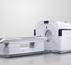
July 25, 2024 — Positron Corporation, a leading molecular imaging medical device company offering PET & PET-CT imaging ...
July 25, 2024
June 18, 2024 — Positron Corporation, a leading molecular imaging medical device company offering PET and PET-CT ...
June 18, 2024Sponsored Content

Feature | Cardiac Imaging
Cardiac positron emission tomography (PET) is growing in popularity among cardiologists because it provides the ability ...
March 05, 2024
June 14, 2024 — Positron Corporation, a leading molecular imaging medical device company offering PET and PET-CT(Positro ...
June 14, 2024
April 23, 2024 — CDL Nuclear Technologies, a pioneer in advanced diagnostic solutions, is proud to announce the launch ...
April 23, 2024
Cardiac positron emission tomography (PET) is growing in popularity among cardiologists because it provides the ability ...
March 05, 2024
October 5, 2023 — Jubilant DraxImage Inc., a wholly-owned subsidiary of Jubilant Pharma Limited, has entered into an ...
October 05, 2023![Phase III clinical trial of [18F]flurpiridaz PET diagnostic radiopharmaceutical meets co-primary endpoints for detecting Coronary Artery Disease (CAD)](/sites/default/files/styles/content_feed_medium/public/Screen%20Shot%202022-09-13%20at%203.30.13%20PM.png?itok=2w6OoNd6)
September 14, 2022 — GE Healthcare and Lantheus Holdings Inc have announced that the recent Phase III clinical trial of ...
September 14, 2022
September 7, 2022 — The American Society of Nuclear Cardiology (ASNC) and three partner societies have come together to ...
September 07, 2022
July 11, 2022 – The American Society of Nuclear Cardiology (ASNC) and the Society of Nuclear Medicine and Molecular ...
July 11, 2022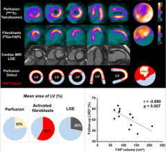
June 15, 2022 — Poor functional outcomes after a heart attack can be predicted with a new PET imaging agent, 68Ga-FAPI ...
June 15, 2022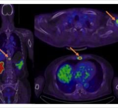
July 13, 2021 — In a recent blog, the American Society of Nuclear Cardiology (ASNC) reported that Humana, one of the ...
July 13, 2021