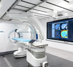
Cedars-Sinai investigators are evaluating novel AI approaches and exploring how these models can be integrated in standard patient care. Image courtesy of Cedars-Sinai
October 31, 2022 — Several recent discoveries show that the accuracy of diagnosing coronary artery disease and predicting patient risk is improved with the help of artificial intelligence (AI) models developed by scientists in the Division of Artificial Intelligence in Medicine at Cedars-Sinai.
These advances, led by Piotr Slomka, PhD, director of Innovation in Imaging at Cedars-Sinai and a research scientist in the Division of Artificial intelligence in Medicine and the Smidt Heart Institute, make it easier to detect and diagnose one of the most common and deadly heart conditions.
Coronary artery disease affects the arteries that supply the heart muscle with blood. If not treated, it can lead to a heart attack or other complications like arrhythmia or heart failure.
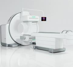
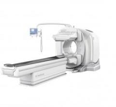
The condition, which affects roughly 16.3 million Americans aged 20 and older, is commonly diagnosed using single photon emission computed tomography (SPECT) and computed tomography (CT) imaging. However, the images generated during the scan aren’t always easy to read.
“We’re continuing to show that AI can improve the quality of images and reveal more information, which makes for more accurate disease diagnoses,” said Slomka, who is also a professor of Medicine and Cardiology and senior author of three studies that were recently published involving AI improving cardiac imaging.
Using AI to Improve Heart Imaging
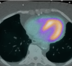
The first study, published in The Journal of Nuclear Medicine, uses AI technology for heart imaging that helps improve the diagnostic accuracy of SPECT imaging for coronary artery disease by advanced image corrections.
In SPECT imaging, it is important to have attenuation correction, which helps to reduce artifacts in heart images, making them easier to read and more accurate. However, it requires an additional CT scan and expensive hybrid SPECT/CT equipment, which is essentially two scanners in one.
While CT attenuation correction has been shown to improve diagnosis of coronary artery disease, it is currently only performed in a minority of scans due to additional scan time, radiation and limited availability of this expensive technology.
To help overcome these obstacles, Slomka and his team developed a deep-learning model called DeepAC to generate corrected SPECT images without the need of expensive hybrid scanners. These images are generated by AI techniques similar to those used to generate “deep-fake” videos and are able to simulate high-quality images obtained by hybrid SPECT/CT scanners.
The team compared the diagnosis accuracy of coronary artery disease using the non-corrected SPECT images—which are used in most places today—advanced hybrid SPECT/CT images, and new AI-corrected images in unseen data from centers never used in DeepAC training.
They found that AI created images that were near the same quality and allow similar diagnostic accuracy as the ones obtained with more expensive scanners.
“This AI model was able to generate DeepAC images in a fraction of a second on standard computer software and could readily be implemented in clinical workflows as an automatic pre-processing step,” Slomka said.
Predicting Major Adverse Cardiac Events
In the second study, published in the Journal of American College of Cardiology: Cardiovascular Imaging, the team demonstrated that deep learning AI makes it possible to predict major adverse cardiac events, such as death and heart attacks, directly from SPECT images.
Investigators trained the AI model using a large multinational database that included five different sites with over 20,000 patient scans. It included images depicting heart perfusion and motion for each patient.
The AI model incorporates visual explanations for the physicians, highlighting the images with the regions that are contributing to high risk of adverse events.
The team then tested the AI model in two separate sites with over 9,000 scans. They found the deep learning model predicted patient risk more accurately than the software programs used currently in the clinic.
“In the first study, we were able to demonstrate that AI can be used to perform important image corrections without the need for expensive scanners,” Slomka said. “In the second, we show that the existing images can be utilized in a better way—predicting patient risk of heart attack or death from images, and highlighting the heart features which indicate that risk, to better inform clinicians about coronary artery disease.”
“These findings represent proof of principle for how AI can augment clinical diagnostics," said Sumeet Chugh, MD, director of the Division of Artificial Intelligence in Medicine. “AI-powered enhancements to SPECT imaging have the potential to improve the accuracy of diagnosing coronary artery disease, while doing it significantly faster and cheaper than current standards.”
Reducing Bias in AI Models
The third study, published in the European Journal of Nuclear Medicine and Molecular Imaging, describes how to train an AI system to perform well in all applicable populations—not just the population the system was trained on.
Some AI systems are trained using high-risk patient populations, which can cause systems to overestimate the disease probability. To ensure that the AI model works accurately for all patients and reduce any bias, Slomka and his team trained the AI system using simulated variations of patients. This process, called data augmentation, helps to better reflect the mix of patients expected to undergo the imaging tests.
They found the models that were trained with a balanced mix of patients more accurately predicted the probability of coronary artery disease in women and low-risk patients, which can potentially lead to less invasive testing and more accurate diagnosis in women.
The models also led to lower false positives, suggesting that the system can potentially reduce the number of tests the patient undergoes to rule out the disease.
“The results suggest that enhancing training data is critical to ensuring that AI predictions more closely reflect the population that they will be applied to in the future,” Slomka said.
The investigators are now evaluating these novel AI approaches at Cedars-Sinai and exploring how these can be integrated in clinical software and how they could be used in standard patient care.
The research was supported in part by the National Heart, Lung, and Blood Institute.
For more information: www.https://www.cedars-sinai.org/
 June 11, 2024